The mouth hardly opens
A narrow gap remains between the teeth. In a series of 521 fused joints, the mouth opened an average of 5.4 mm before surgery.
 Discuss your case
Ankylosis of the temporomandibular joint is a fusion of the jaw joint with the skull by bone or scar tissue, due to which the mouth barely opens or does not open at all. It is treated with surgery, and after it, regular development of the jaw is important so that the fusion does not return.
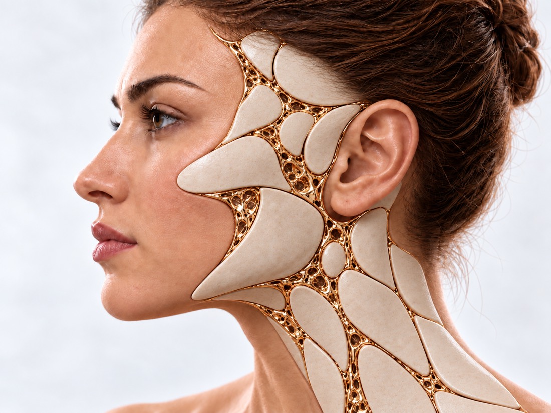 CONCEPTUAL VISUALIZATION
The temporomandibular joint is the joint in front of the ear where the lower jaw moves when you open your mouth, chew, and speak. With ankylosis, the head of the jaw grows with the articular fossa of the skull by bone or dense scar (fibrous) tissue.
This makes it difficult to chew, speak and brush your teeth. And in an emergency situation, the fused joint becomes dangerous: it is difficult for doctors to ensure breathing when the mouth does not open.
Bone ankylosis occurs more often than fibrous ankylosis, and unilateral - more often than bilateral. If the joint grew during childhood, the jaw on this side grows worse: asymmetry, a small lower jaw appear, and sometimes breathing stops during sleep.
This page is about a fused joint. If the head of the joint is gradually destroyed and the bite changes, and the mouth opens at the same time, this is another condition.
This is general information. The doctor will tell you what will suit you after the examination.
The main symptom is that the mouth opens less and less. In children, facial and breathing changes are added to it over time.
A narrow gap remains between the teeth. In a series of 521 fused joints, the mouth opened an average of 5.4 mm before surgery.
You have to eat bland food, it is difficult to take care of your teeth and mouth.
If ankylosis occurred in childhood, the jaw is underdeveloped, the chin shifts or "runs" back.
Asymmetry of the face and jawsLoud snoring, pauses in breathing, snoring or choking in sleep are possible signs of obstructive sleep apnea, which can accompany ankylosis with an underdeveloped jaw.
Most often, ankylosis is the result of an injury, often in childhood. The authors of one of the studies suggest that ankylosis is more common where little is known about it and a fresh injury is initially treated incorrectly.
The most common cause of ankylosis. In a survey of 1,607 children at one specialist center in India, 128 cases of ankylosis were found, and the main cause was trauma during childbirth or during childhood.
Fractures of the condylar processThe second most frequent cause after trauma.
Joint fusion is also a consequence of autoimmune diseases.
In a series of 28 children with congenital facial defects, ankylosis in 9 was congenital, in 16 it occurred after previous operations on the jaw, in 3 — after infection.
In a series of 28 patients (mean age, 51.8 years), who received an artificial joint, ankylosis was after trauma in 39%, after arthrosis in 32%, and congenital in 25%.
Chronic ankylosis usually does not require emergency care - it is treated as planned. But due to the fused joint, it is more difficult to help a person if he is suffocating, so you should warn doctors about ankylosis in advance.
Immediately - to the surgeon who operated, and if it does not work - to the waiting room: after the operation, the pain increases, the wound becomes red, swollen or pus is released from it, a fever appears. The same day to the doctor: the mouth suddenly stopped opening or the jaw jammed; you cannot eat or drink; after a blow to the chin or face, it hurts in front of the ear and the mouth does not open completely - especially in a child. Planned: the mouth opens less and less over the months; the child snores loudly and stops breathing in his sleep; the jaw grows asymmetrically.
When the mouth began to open worse, were there injuries or infections. The doctor measures how many millimeters the mouth opens.
A CT scan shows where and how the joint has fused—with bone or scar tissue—and how the jaw has changed.
If there is snoring and breathing pauses, especially in a child with a small jaw, check breathing in sleep.
The mouth does not open, so they plan in advance how to safely ensure breathing during anesthesia.
The goal of treatment is to restore jaw movements, correct facial deformity, reduce pain, and prevent re-union. All of the methods that the reviews compare are surgical; exercises complement surgery, not replace it.
The fused area is removed to allow the jaw to move again.
When the head is removed together with the fusion, the branch of the jaw is restored.
Ankylosis of the temporomandibular joint is treated at Dr. Valikhnovskyi's Institute of Surgery in the "New Face" direction - in the "Lower Jaw and Joint" area, along with fractures and reconstruction of the jaws.
Plastic surgeon, candidate of medical sciences, founder of Dr. Valikhnovskyi Institute of Surgery.
Short answers. What is right for you, the doctor will tell you during the consultation.
The main treatment is surgery: all the methods that the reviews compare are surgical. Exercises and devices for developing the jaw are needed after it to maintain the result.
Yes, this is the main risk. In a series of 521 joints, ankylosis returned in 8.82%, in a meta-analysis of own grafts - in about 8%. That is why exercises after surgery are so important.
Depends on the method and exercises. In a series of 521 joints — an average of 36.9 mm after a year; according to a meta-analysis of transplants, the mouth opened 27–31 mm more than before surgery.
There is no unequivocal answer. According to the 2019 meta-analysis, the results are similar. A child's rib can grow unpredictably, so a prosthesis is more often considered for adults. The method is chosen based on age, CT and the condition of the jaw.
The doctor determines the terms individually. Ankylosis interferes with the growth of the jaw and can disrupt breathing in sleep, so the plan is made taking into account growth, breathing and bite.
If the jaw is underdeveloped, one release of the joint may not be enough: the jaw is pushed out or aligned - at the same time or at a later stage.
Because the mouth does not open, and in an emergency situation or during anesthesia, it is more difficult to ensure breathing. This is planned in advance.
The figures on the page are from these sources.
Send CT scan if available and photo of face. The doctor will tell you what can be done and where to start.
Leave your contact details and briefly describe the situation. A coordinator will contact you to agree on the next step.