
FACE / RECONSTRUCTION

Facial reconstruction.

We restore facial form, symmetry, expression and function after trauma, wounds, burns, tumour removal and congenital changes.

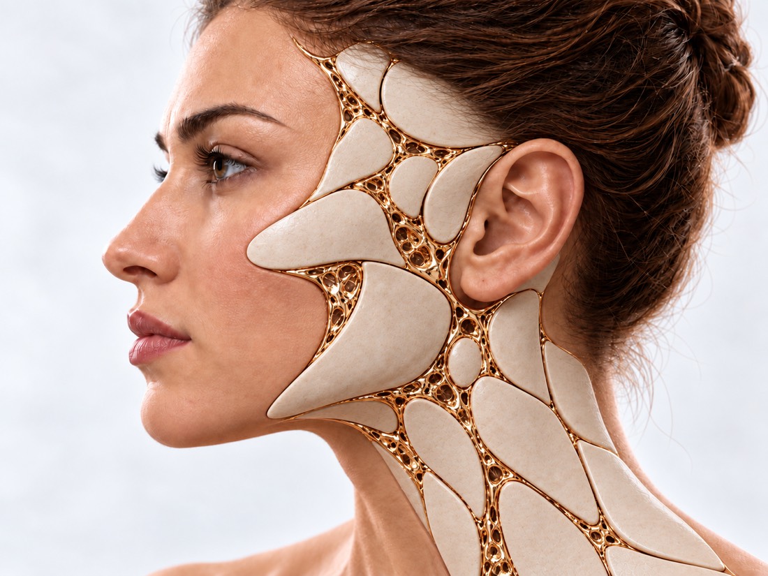
Conceptal visualization directly "New Face": face with gold lines of reconstructionFIG. 02 / CONCEPTUAL VISUALIZATION

Just had an injury?

First — emergency care, then do not delay consulting a surgeon: fresh fractures are easier to treat until the bone has grown.

103 or directly to the reception room if:

  • A person was unconscious, a judge, unusual sleepiness or language abuse;
  • double vision, reduced vision, eye pain or impaired eye movement;
  • There was vomiting;
  • The forced haemorrhage does not stop in the nose or purple edema;
  • A transparent watery liquid flows from the nose or ear;
  • A big wound, or a wound, glass or wreckage.

Burn of the face.

Cool the burn with cool running water for 20 minutes, do not apply creams or oil. Hoarse voice, boiling in your mouth, burnt hair on your nose or difficulty breathing — just call 911: swelling of the airways may increase.

Part of the face does not move after the wound.

On the same day, the surgeon is best able to sew the facial nerve as early as possible.

What is
facial reconstruction.

A person's recommendation is operations that restore bones, soft tissue and facial mimics.

Modern facial reconstruction is a combination of microsurgery, digital modeling and regenerative technologies. Today the philosophy of surgeons has evolved from “just close the wound” to restoring “like for like”. The main task — to preserve expression, chewing, swallowing, language and restore as much as possible facial features after severe mine-explosion injuries, oncological operations or accidents.

Reasons to seek treatment include changes in facial form or function: a sunken eye or double vision, facial numbness, limited mouth opening, altered bite, a residual bone or soft-tissue defect, or a scar that pulls on the eyelid or lip.

The specific indications, method and expected result are determined individually by the doctor after consultation and necessary examinations.

From damage
to recovery.

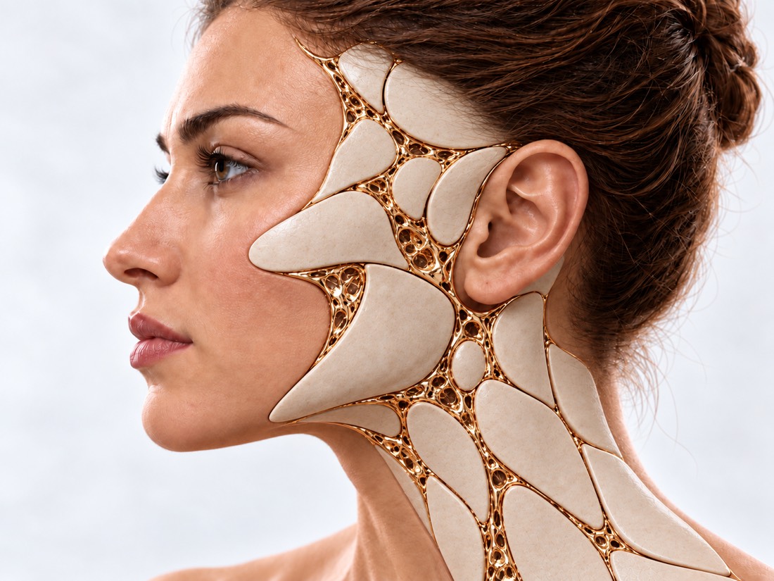
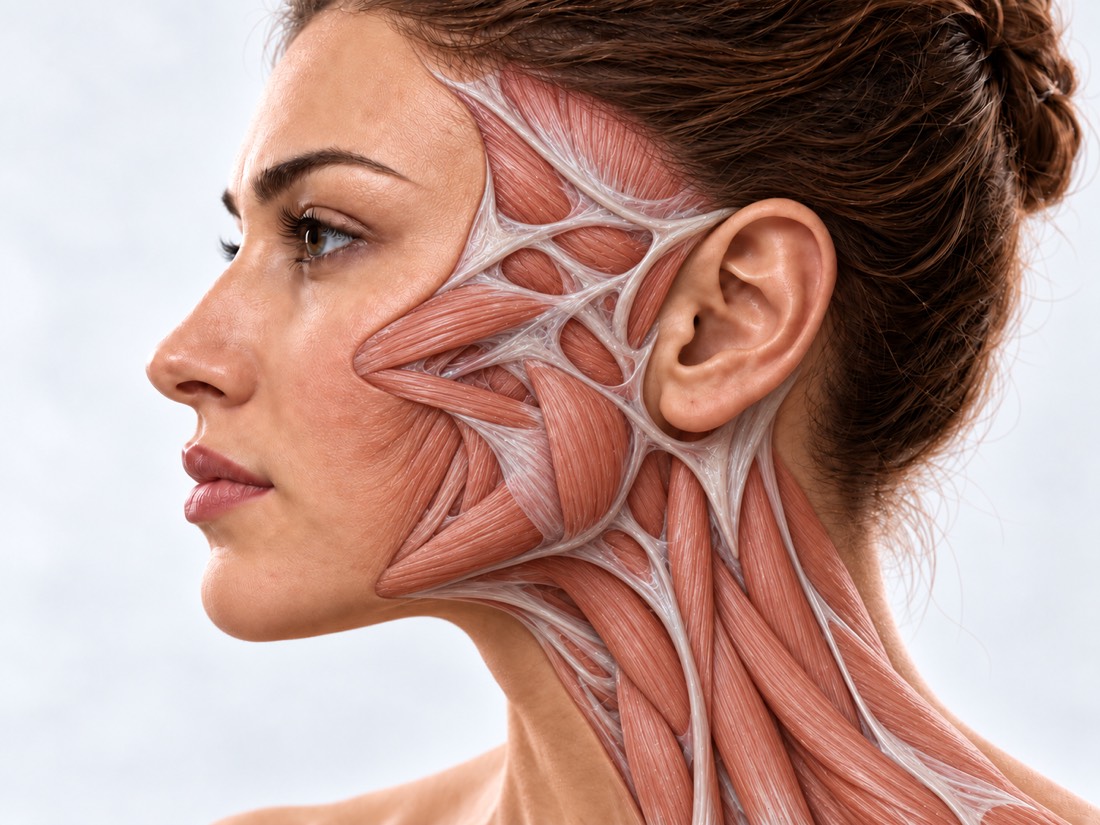
Move the slider. Open anatomic layer means damaged tissue, gold means the reconstruction performed.

The artist is direct, not a picture of the patient.

Golden facial reconstruction in conceptual visualization Opening muscles and facial tissue in conceptual anatomical visualization BACKGROUNDRECONSTRUCTION
FIG. 02.1 / CONCEPTUAL VISUALIZATION, NOT PACENT PHASE

What can be
restored.

Six situations that are most common.

01

Fractures and deformities after injury.

Twice, orbit, jaws, nose, frontal puzzle: fresh fractures and bones that have grown wrong.

How it is treated

Whether surgery is needed is determined by the displacement of fragments, symptoms and CT: some fractures are treated without surgery or with closed reposition. When surgery has to be done, the fragment is aligned and fixed with titanium plates; if indicated, an implant is used to restore the orbital wall. If bones have grown together with the deformation, they are cut off and replaced (osteotomia) or contoured with implants or grafts.

Post-traumatic deformation of the face
02

Firearm and landmine injuries.

At the same time, affected bones, skin, muscles, and nerves remain debris in the tissue.

How it is treated

A fresh wound is first treated according to the rules of emergency care: breathing, stopping bleeding, cleaning the wounds. Reconstruction is planned after CT, condition of the wound and general state of patient, often in several stages. Whether to remove debris, the surgeon decides after a CT.

Surgery of firearms injuries
03

Defect after tumor removal.

There are fewer tissue left after the skin, jaw, or soft tissue tumor is removed.

How it is treated

Small and medium defects are covered with local flaps from the face and neck – they are closest in color and texture. Large, along with bone, by microsurgical flap. Reconstruction is often done in one operation with tumor removal if the edges of the incision are clean; otherwise after histology.

Surgery of soft tissue
04

Scars from wounds and burns.

The ribs draw their eyelids and lips, change the contour of their faces, prevent them from closing their eyes and opening their mouths.

How it is treated

The scars are excised and covered with local tissue; for large areas an expander is used. Keloid scarring may recur after simple excision, so it is treated in a combination: post-excision injection into the scar or radiation therapy as prescribed by the doctor.

Recommendation for burns
05

Paralysis of the facial nerve.

The angle of the mouth goes down, the eye doesn't close completely, the face loses symmetry.

How it is treated

After a closed injury, nerve function often recovers on its own. The cut nerves are best sutured in the first few days; surgery is possible for about one year. In prolonged paralysis, movement can be partially restored by muscle transplantation and static operations improve symmetry at rest.

Paralysis of facial nerve
06

Congenital facial changes.

Splits, maldevelopment of half the face, ear shell or jaw.

How it is treated

Children tend to have some form of treatment in view of the growth and development of speech, bite and vision.

Congenital defects surgery

What is restored
and with what.

Surgeon material is designed for each defect separately and explains the risk of each option.

AreaRemediationThan
Orbit and ForeverOrbits, eyes and eyelidsIndividualized orbit implant, chrome transplant
Silk and middle zoneIt's like a complex, top jaw.Titan plates, osteottomy
Lower jaw and jointbody and angle of jaw, joint, biteplates, a fibular bone flap and a joint implant
NoseEngine, partition, frameBrisket or ears — New nose
Forehead and cranial vaultBone defects, elbow contourBone, implant.
Skin and soft tissuescarring and defects after wounds, tumors and burnsLocal flaps, tissue expanders, free flaps and lipofilling.

How faces are
restored.

The algorithm depends on the size, location of the defect and the attraction of deep structures.

01

Virtual Planning (VSP) and 3D printing.

It's the foundation of modern jaw-faced reconstruction that reduces errors during surgery.

Mirror CT modelling
Using computed tomography, the intact half of a patient's face is "mirrored" in a special program, creating an accurate three-dimensional model for restoring the damaged side.
Individual implants (PSI)
Medical grade titanium or PEEK biopolymer is 3D-printed or milled into individual plates, orbital grids or jaw fragments that accurately replicate the patient’s anatomy.
Surgical templates
During the operation, the surgeon applies them to the bone in order to make cuts at a specified angle.
02

Free microsurgical flaps

Large bone and soft tissue defects replace tissue from other parts of the body by sewing the vessels under the microscope.

Tiny bone (Fibula flap)
A fragment of the tibia bone, together with blood vessels and skin, is used to recreate a lower or upper jaw. The bone is cut into 3D templates, bent in the shape of a jaw, fixed with titanium, and the blood veins are sutured to the cervical arteries and vein.
Radial forearm flap
A thin, flexible skin flap used to reconstruct the tongue, lips, cheeks, oral mucosa and floor of the mouth.
Antherolatheral hip strap (ALT) and too small ALT
In the ultra-thin version, the surgeon removes most of the subcutaneous fat leaving a thin layer of skin with fat and blood vessels – this reduces the flap volume and mask effect on the face.
03

Local flaps and expander.

Small and medium defects are better to cover their faces and necks: they are the closest in color and texture.

Facial artery perforator flaps (FAP)
The surgeon uses ultrasound dopplerography to locate the small vessels (perforants) that flow from the facial artery and moves a flap of skin on this thin vascular leg. This allows wounds to be closed without rough tensioning or distorting lines of mouth or eyes.
Clusters of Surrounded Zones
The nasolabial, cervical and frontal flaps. Thanks to the rich blood supply of the face these flap are well implanted. For a nose flab — on the page "New Nose".
Tissue dermotenasia (expander)
A silicone balloon is inserted into a healthy area next to the defect – on the cheek, neck, forehead or hairline of the head – and gradually filled with physiosolution over 1-3 months. During the second operation, the expander is removed and excess healthy skin similar in color is moved onto the facial defect.
04

Restoration of facial expressions.

If the facial nerve or mimical muscles are damaged, the face loses symmetry and mobility. Modern surgery can partially restore a smile thanks to dynamic reinervation.

Cross-facial plastic nerve
A nerve graft — most often a sular nerves from the shin — connects a branch of a healthy facial nerver on the opposite side to the affected side and transmits mimic impulses.
Gracilis muscle transfer
A segment of the gracilis muscle from the inner thigh is transplanted to the face. Its vessels are connected to local vessels, while its nerve is connected either to the masseteric nerve or to a cross-facial nerve graft. When the patient clenches their teeth or tries to smile, the transplanted muscle contracts and lifts the corner of the mouth.
05

Regenerative surgery and lipofilling.

Own fat is used to supplement contours after large reconstructions and as an auxiliary method for the scar.

Lipofield: Microfat & Nanofat grafting
The patient's own fat is cleaned and inserted with thin cannulas into the areas of asymmetry. Nanofat contains almost no live fat cells, but it does contain stromal-vascular fraction with stem cells. When introduced superficially under the skin, it may improve the texture, color and softness of scars, especially after burns and radiation therapy; this has been best confirmed in short term.
06

Alloplasty of the face.

A layer of options for the most difficult cases where normal reconstruction cannot restore a person's function and appearance.

Donor's facial transplant
A patient is transplanted a complex of facial tissues (skin, subcutaneous fat, muscles, nerves, sometimes bone) from a post-mortem donor. This requires lifetime immunosuppression with serious risks: rejection, infections and tumors. Only dozens of such operations have been performed worldwide.

What happens
during reconstruction.

Gold marks the tissue or model used to reconstruct the defect.

01

Mirror planning.

Virtual planning: half of the person ' s health affects damage Healthy Side mirror copy outline and bones plan defect After injury or tumor

During CT scans, the healthy half of the face is mirrored on the damaged one. This results in a plan for the contour and bones that are used to print templates and individual implants.

02

Jaw made of the femur bone.

The small bone fragment is sprayed into segments and formed by the lower jaw. Capture receptacle 1 tibia bone retained jaw parts Seeds — To the Neck 2 night jaws

A fragment of the tibia bone with its own vessels is cut into segments using a 3D template, folded into jaw shape and fixed with titanium. The flap vessel is sutured to the neck — the bone lives in a new place.

03

Restoration of a Smile

Dynamic resuscitation: Nervous transplant on the healthy side and muscle transplant on the affected side Healthy facial nerve M. hip muscle the angle of the mouth. Neutrons Transplant From the ankle

A nerve graft from the femur is inserted from a branch of a healthy facial nerves to the affected side. The transplanted muscle from thigh is attached to an angle of the mouth: when it contracts, the angle rises.

The schemes are simplified and not scaled up.

When to do
surgery.

  1. Fresh fracture

    Bones are easier to deliver until they grow up; sometimes the operation is postponed for a few days until the swelling is reduced.

  2. Wound

    The term refers to wounds and patients; reconstruction often begins.

  3. Rubles

    Planned – after the scars have matured, when they become paler and softer. Earlier – if the Scar prevents you from closing your eye or opening your mouth.

  4. Pulse nerve and tumor

    The cut nerve is sutured in the first few days. After tumor removal, reconstruction is often done in one operation if the tumour has been completely removed.

How the recovery
is going.

Terms depend on the number of transactions.

First weeks of pregnancy

Swelling and bruising are greatest in the first days, with major swelling passing within a few weeks. After orbit surgery, the twins due to swelled usually gradually decrease.

Up to a year-two.

The rubles are ripe and glowing up to a year or two.

After surgery on the jaws.

Sometimes food is only soft or liquid. After surgery on the orbit, jaw or upper jaw do not eat until your surgeon allows you to. Do not smoke: this worsens healing.

When to call 103?

Difficulty breathing or swallowing, swelling in the mouth or neck builds up quickly; bleeding does not stop; after surgery on the orbit eye pain increases, eye is suspended or vision deteriorated. Temperature of 38 °C and above or pus — to the surgeon the same day.

Rehabilitation of
the face.

Which brings back movement and function.

01

Paralysis of the facial nerve.

Exercises for the Face
Learn more

In a review of 7 randomized trials for peripheral facial nerve paralysis, physical therapy could half the rate of patients not recovering (relative risk 0.51; 4 studies, 396 participants) and improve Sunnybrook score but evidence quality is low. An updated systematic review on Bell’s palsy reinforces conclusions about early exercise benefits and adds evidence on their value in chronic cases.

Sinkinesia
Learn more

Synkinesies are involuntary associative movements when a nerve after paralysis recovers incorrectly, for example the eye is tensed during smile. An international group of experts recommends starting with biofeedback training followed by botulinum toxin; surgery is left for individual cases. Botulinum Toxin improved scores on the synkinesis questionnaire but there are no single dose recommendations.

Paralysis after surgery
Learn more

After surgery (mostly on the eardrum), physical therapy is likely to be beneficial and not harmful, although due to the diversity of studies there are no definitive conclusions (review of 15 studies, 313 patients).

02

Opening the mouth, tongue and chewing.

Trism
Learn more

Trismia – limited mouth opening. After treatment of head and neck cancer, exercise combined with a jaw-development device increased the openness of the brow; prophylactic exercise failed to prevent trismias (13 randomized trials, 733 patients).

Language and chew
Learn more

After bone flap reconstruction of the lower jaw, tongue was restored best and chewing worst (257 patients, follow-up on average over 3 years).

How much
does it cost.

The value of the surgery is referred to by the surgeon after examination and CT:

  • Countries and volume of transactions;
  • Material: its fabric, standard or individual implant;
  • Periods;
  • The duration of the infirmary and the days in the hospital.

From the first
call.

  1. Conversations and materials

    You describe the request and, if you have it, you provide the results of the previous examination and treatment.

  2. Consultation and evaluation

    The doctor determines what situation we are in, what circumstances we need, what we need, and what his limitations are.

  3. Reorganization

    It discusses the next steps, the sequence of phases and the issues of reconstruction.

  4. You don't live in Kiev or abroad?

    Patients come from all over Ukraine and abroad.

What is worth
discussing?

01

What happened and when?

02

What's broken: chew, eye, mimic, speech?

03

What kind of surgeries have you had?

04

Is there a C.T., a discharge and a photo of the face before the injury?

05

What chronic diseases and medications?

06

What's the most important thing?

Frequently Asked
Questions.

The short answers are that what suits you is what the doctor will tell you.

Will there be scars?

Access to the bones is made in hidden places where possible: from the side of the mouth, along the inside surface of an eyelid, behind a hairline. Most scars lighten over time; keloid scarring requires separate treatment.

When is it best to do post-traumatic reconstruction?

Fresh fractures are easier to fix until the bone has grown incorrectly, so you should not delay with consultation. If the Bone has already growt in a displacement, it is corrected plannedly.

When can we operate on the scars?

While the scar is ripening, it can be treated without surgery: silicone, pressure, massage as prescribed by a doctor. Early operation is performed if the Scar prevents you from closing your eye or opening your mouth.

Should the debris be removed from the face?

Not always. Wood, plant or plastic fragments are usually removed: they cause infection. Metal or glass without symptoms near important structures sometimes remain and are observed. The decision is made after examination and CT scan.

Can you turn a smile with a facial nerve palsy?

Fresh nerve rupture is sutured in the first days; with prolonged paralysis, movement can be restored by transplanting a muscle from the thigh and static operations improve symmetry at rest. The method is chosen individually.

Can dental implants be installed after the jaw reconstruction?

For example, a bone transplanted from the tibia can be implanted with dental implants to restore chewing. The timeframe is determined after the bone has survived.

What tests should be conducted before the operation?

The main one is a CT scan of the facial skull; in case of orbital injuries, an ophthalmologist’s examination is mandatory. Before general anaesthesia — tests and an evaluation by an anesthesiologist.

Sources

15 NHS, AO Foundation and peer reviews
ShowTurn to Head
  1. Head injury and concussionNHS (United Kingdom)nhs.uk/conditions/head-injury-and-concussion ↗
  2. Burns and scaldsNHS (United Kingdom)nhs.uk/conditions/burns-and-scalds ↗
  3. Accuracy of virtual planned surgery versus conventional free-hand surgery for reconstruction of the mandible with osteocutaneous free flapsInternational Journal of Oral and Maxillofacial Surgery, 2020doi:10.1016/j.ijom.2020.02.018 ↗
  4. The Facial Artery Perforator Flap for Reconstruction of Facial Defects: Surgical Pearls and Clinical SeriesJournal of Craniofacial Surgery, 2024doi:10.1097/SCS.0000000000010025 ↗
  5. Donor nerve selection in free gracilis muscle transfer for facial reanimation. A systematic review and meta-analysis of clinical outcomesJournal of Plastic, Reconstructive & Aesthetic Surgery, 2023doi:10.1016/j.bjps.2023.04.014 ↗
  6. Effects of Nanofat in Plastic and Reconstructive Surgery: A Systematic ReviewPlastic and Reconstructive Surgery, 2024doi:10.1097/PRS.0000000000010905 ↗
  7. Outcomes in facial transplantation - a systematic reviewBritish Journal of Oral and Maxillofacial Surgery, 2024doi:10.1016/j.bjoms.2024.02.008 ↗
  8. Orbital floor fracture: open reduction with or without internal fixationAO Surgery Reference, AO Foundationsurgeryreference.aofoundation.org/cmf/trauma/midface/orbit-floor/open-reduction-with-or-without-internal-fixation ↗
  9. Physical therapy for peripheral facial palsy: A systematic review and meta-analysisAuris Nasus Larynx, 2024doi:10.1016/j.anl.2023.04.007 ↗
  10. Physical therapy for facial nerve paralysis (Bell's palsy): An updated and extended systematic review of the evidence for facial exercise therapyClinical Rehabilitation, 2022doi:10.1177/02692155221110727 ↗
  11. Pathogenesis, diagnosis and therapy of facial synkinesis: A systematic review and clinical practice recommendations by the international head and neck scientific groupFrontiers in Neurology, 2022doi:10.3389/fneur.2022.1019554 ↗
  12. Botulinum toxin A treatment in facial palsy synkinesis: a systematic review and meta-analysisEuropean Archives of Oto-Rhino-Laryngology, 2023doi:10.1007/s00405-022-07796-8 ↗
  13. Physical Therapy for Iatrogenic Facial Paralysis: A Systematic ReviewJAMA Otolaryngology–Head & Neck Surgery, 2020doi:10.1001/jamaoto.2020.3049 ↗
  14. Exercise therapy for cancer treatment-induced trismus in patients with head and neck cancer: A systematic review and meta-analysis of randomized controlled trialsRadiotherapy and Oncology, 2020doi:10.1016/j.radonc.2020.08.024 ↗
  15. Long-term functional outcomes of vascularized fibular and iliac flap for mandibular reconstruction: A systematic review and meta-analysisJournal of Plastic, Reconstructive & Aesthetic Surgery, 2021doi:10.1016/j.bjps.2020.10.094 ↗

Emergency signs and first aid are for NHS, while others are peer reviews of methods described on the page.

VALIKHNOVSKI SURGERY PARTNERS

Your case
deserves attention.

Start consulting to see what questions and materials are needed for the evaluation.

Call Send a request