BODY / COMPREHENSIVE CARE

Comprehensive body reconstruction.

When several body areas are affected by multiple wounds, extensive burns or necrosis, we plan reconstruction as one sequence of stages.

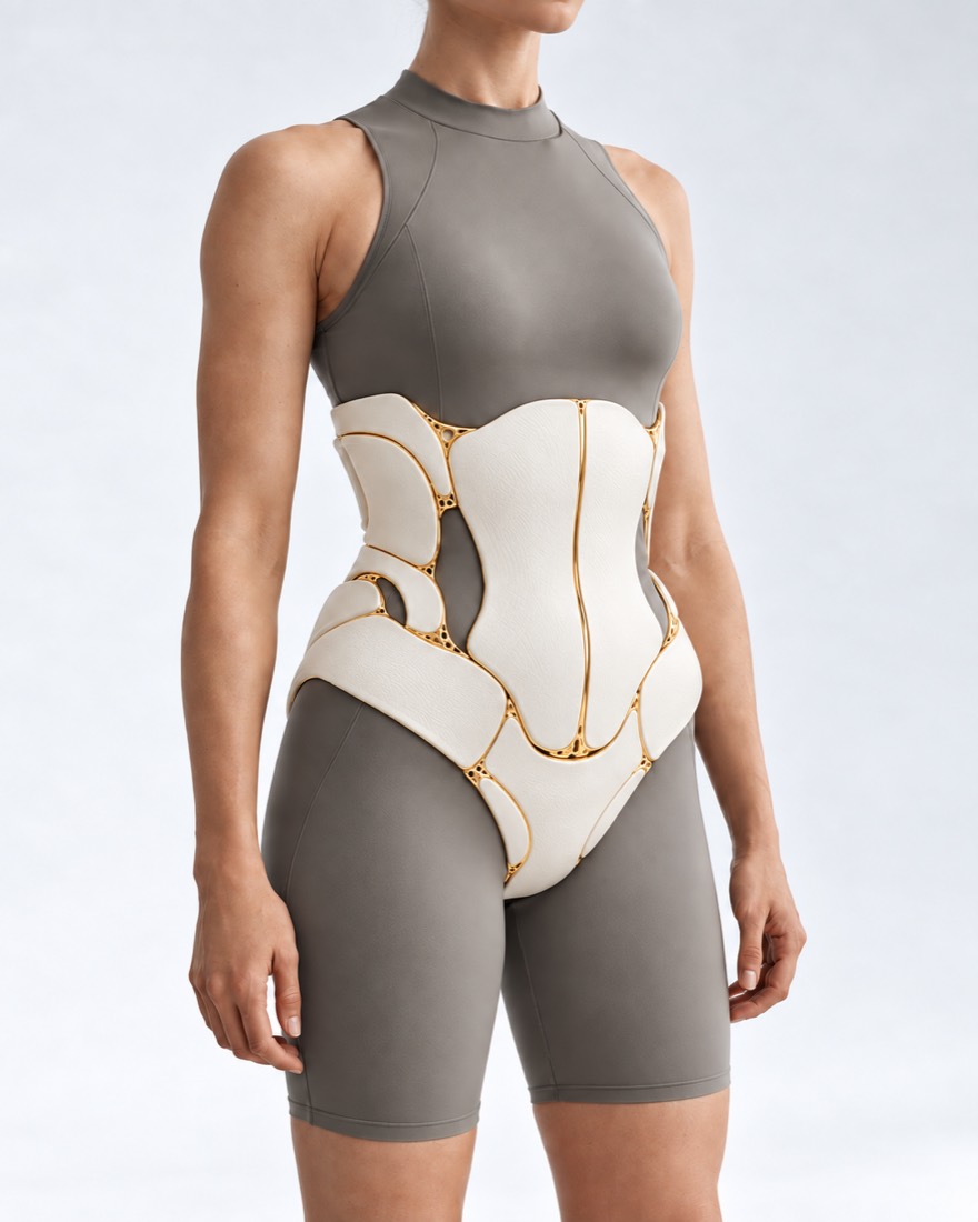
Conceptal visualization directly "New Body": a torso with open anatomical layers and gold linesFIG. 12 / CONCEPTUAL STAFFING

What is
complex reconstruction.

Complex body reconstruction is a recovery plan where several areas are affected at once and treated in the correct sequence.

Whole body reconstruction is not a single operation, but a multi-stage treatment that combines reconstructive microsurgery, burn management, traumatology and individual planning based on CT data. It is used for severe lesions of several areas: after mine explosion injuries, large burns (usually more than 30% of the body surface), cancer operations or necrotizing fasciitis.

They come after discharge from the hospital or burn center, when there are many wounds, scars and impaired functions, and it is unclear where to start.

The specific indications, method and expected result are determined individually by the doctor after consultation and necessary examinations.

When a joint
plan is needed.

Six situations that are most common.

01

Multiple injuries.

Several areas have been affected since the mine injury: faces, limbs, torso.

How it is treated

Compile a single plan: what threatens function — first, then closure of wounds, bones, scars. Separate zones lead profile directions .

Surgery of firearms injuries
02

Severe Burns.

Glasses on most of the body, little healthy skin for transplantation.

How it is treated

Wounds are closed with mesh and Meek grafts, cultured cells, dermal matrices; then — staged reconstruction of scars.

Recommendation for burns
03

After necrotizing fasciitis fasciitidis.

After a serious infection, large areas of skin and tissue were removed.

How it is treated

Once the infection is controlled, wounds are managed with negative-pressure therapy and closed with grafts or flaps; body contours are then restored.

Flankless fascitis
04

Lymphedema after injury Lymphoedema following injuries

A hit in the lymphatic path leads to a fatherland.

How it is treated

First, compression and lymphatic drainage; if indicated — lymph venous anastomosis: small lymphoid vessels are connected to the veins under a microscope.

Post-traumatic lymphadem
05

Contour deformations.

Tight scarring, short volume, scarring weights on the torso and limbs.

How it is treated

Rubles are planted and plasticized by local tissue; they are filled with fat.

Body perturbation
06

Amputations

The end is lost before the prosthetics.

How it is treated

The coccyx is prepared for the prosthesis: nerve transfer (TMR) to reduce phantom pain, if indicated — osteointegration. More information — in the direction «Bionic prostheses».

Bionic limb prostheses

What is the
reconstruction.

Methods used in modern recovery surgery.

01

Stage tactics of Damage Control and NPWT-systems.

In the case of critical political vascular or large-scale burns, the patient may not survive many hours of reconstruction at once.

Damage Control Surgery
At the first stage, surgeons perform only life-saving interventions – stopping bleeding, stabilizing fractures with external fixation devices and removing dead tissue.
Vacuum closing (NPWT)
Large wound surfaces are covered with vacuum systems to drain wounds, stimulate granulation growth and prepare the wounding for closure.
02

Closure of large wounds with skin deficiency.

After almost no healthy skin is left for transplantation, special methods are used.

Meek microdermoplastics
A thin skin graft is cut by a special machine into small squares (about 3 mm) and stretched on a specific corrugated fabric in the ratio of 1:9. This enables to close a large wound with minimal amount of donor skin.
Cultural Ceratorium (CEA)
A tiny bioptat of healthy skin is taken from the patient and layers of its own epidermal cells are grown in the laboratory, which then close wounds.
Piece of wood
Dermal matrices (e.g. Integra) act as an artificial layer of dermis. They grow through the patient's vessels, after which a thin skin graft is placed on top.
03

Microsurgery

To recover lost amounts of muscles, muscles and bones in various anatomical areas.

Free flaps
Tissues from one area of the body are transplanted to another, suturing the valve vessels with local arteries and veins under a microscope. Several valves are sometimes transplanting in one operation by several teams.
Limphovenous anastomosis
With extensive limb injuries, the lymphatic pathways can be damaged leading to chronic swelling (lymphedema). Surgeons suture lymph vessels about 0.3-0,8 mm in diameter with small veins, improving the outflow of lymph from the limber.
04

Bone frame.

Individual implants (PSI)
Based on the CT data, individual titanium implants of chest, ribs, pelvic bones and skull fragments are made for each patient.
Indunal membrane method (Masquelet)
First, a temporary cement spacer is placed in the defect and its own membrane forms around it over several weeks; then the spacers are removed and the cavity in their membrane is filled with bone tissue.
05

Contour reconstruction.

Once the wounds are closed and the bones fold, deformation of the contours, scarring and chronic pain often remain.

Lipotransfer
Fat can soften scars and restore the volume of subcutaneous tissue.
If the limbs are lost
Osteointegration and nerve transfer (TMR, RPNI) " Boonic Prostheses of the extremities ".

How they
watch the klaptem.

The transplanted lymph node is fed through vessels sutured under a microscope. The first two days are crucial: it is important to notice any blood flow problems as early as possible.

01

How often and when?

What the numbers say
In a systematic review of 109 studies (44,031 valves), due to blood flow disorders, 5.8% of the valve was reoperated: 3.7% were saved and 2.1% lost. Among the missing valve, where the cause is indicated, it was most often in veins (59.5%), less frequently in arteries (27.9%).
Decision time
If the inspection was done in the first day, 93.8% of flaps were saved; on the second — 83,3%, on third —12,1%, and later — none. Therefore, the closest monitoring is carried out during the initial 48 hours.
02

Methods of control.

Overview
The colour and temperature of the flap remains the basis for inspection. Instruments only complement the examination, as they can also give false signals.
Doppler implanted
A sensor on the valve vessel continuously transmits a blood flow signal. According to a meta-analysis of 22 studies, sensitivity is 81%, specificity - 97%.
Tissue oxytocin (NIRS)
A light sensor on the skin of the flap measures tissue oxygen saturation. It detected blood flow disorders 82 minutes earlier than clinical signs, on average; it saved 90% of such valves compared to 50%, with examination alone (10 studies, data are still limited).
ICG-angiography
During surgery, a fluorescent dye is injected into the vessels and the camera shows which part of the flap has good blood supply. In head-neck reconstruction this reduced total flaps loss; in breast reconstructions it reduces skin and fat necrosis and reoperations (but not flapes loss); for limbs data are limited and there was no effect; overall quality of evidence low.
03

Rescue of the Flap

Medical leeches
When blood enters the duct but drains poorly, saliva temporarily removes excess. A review of 41 studies found success at 65–85%; however, about half of patients required a transfusion and antibiotic prophylaxis is needed.

How they
close large wounds.

The gold marks the tissue that the defects restore.

01

Meek-Micrografts

Meek method: skin is cut into small squares and stretched up to nine times more Donor skin Wound, area up to ×9

A thin piece of healthy skin is cut into hundreds of small squares and evenly spread over the wound with gaps. The skin grows from each square to close the gap.

02

Lymphovenous Anastomosis

Limphovenous anastomosis: The lymphic receptacle connects to the small vein Limphical receptacle0.3-0.8 mm Small vein Walked under a microscope. Limfa

Lymph, which stagnates through the damaged pathways, is given a bypass into the vein. The suture is made under a powerful microscope: vessels thinner than a millimeter.

03

The sequence of stages:

Step sequence: stabilisation, wound closure, bones and function, contours 1Stabilization 2Close the wound 3Bones, function 4contours Weeksmonths — years

First, life-saving and limb, then wound closure, bones, nerves and function, and only at the end body contours and scarring.

The schemes are simplified and not scaled up.

How to make
a plan.

  1. Overview of all zones

    The surgeon evaluates each area, general state and nutrition; collects CTs and releases.

  2. Priorities

    The first is open wounds, contracts that prevent us from moving and breathing.

  3. Phases at intervals

    There are usually weeks or months between large operations.

  4. Plans

    After each step, the plan is finalized with the patient and the rehabilitation officer.

How the recovery
is going.

Terms depend on size; the plan is individual.

Hospital stay

After the big steps-- in-patient surveillance, dressing.

Rehabilitation

The fluid physical stress between stages is part of the treatment, not the supplement.

Compression

Compression garments are worn over scars for months; for lymphoedema, compression is often long term or permanent.

Nutrition and strength.

Large wounds require protein and energy; healing depends on the general state.

How much
does it cost.

The value depends on the number of areas and stages; the surgeon calls it after verification and planning.

For military and veterans, there are special benefits.

The clinic is responsible for funding charitable foundations for members of the SSO.

From the first
call.

  1. Conversations and materials

    You describe the request and, if you have it, you provide the results of the previous examination and treatment.

  2. Consultation and evaluation

    The doctor determines what situation we are in, what circumstances we need, what we need, and what his limitations are.

  3. Reorganization

    It discusses the next steps, the sequence of phases and the issues of reconstruction.

  4. You don't live in Kiev or abroad?

    Patients come from all over Ukraine and abroad.

What is worth
discussing?

01

Which areas are damaged, and what's the biggest obstacle?

02

What kind of surgeries have been done, and where?

03

Are there open wounds now?

04

Is there a discharge, C.T., a photo?

05

What medications do you take with weight and food?

06

What's the most important thing for you to return first?

Frequently Asked
Questions.

The short answers are that what suits you is what the doctor will tell you.

Where does it start when many areas are affected?

From what threatens function: open wounds, contractures that impede movement or breathing. Appearance and contours — at later stages.

How long does the reconstruction go on?

In general, months or years: between large operations, a break for healing and rehabilitation is required.

Can serious burns be covered if we don't have enough skin?

Yes: skin is stretched with a mesh or Meek method, dermal matrices are used and cell cultured in the laboratory of the patient.

Does the limbs heal after an injury?

If it doesn't work, it's possible that microsurgery is done on lymph vessels.

Do you treat the military?

Yes. Operations for IDF servicemen are carried out by the clinic with funding from charitable foundations; details will be explained by administrator.

Sources

15 sources of communication and research
ShowTurn to Head
  1. Damage control in orthopaedical and traumatologyColombia Médica, 2021doi:10.25100/cm.v52i2.4802 ↗
  2. Outcome comparison of the most commonly employed wound coverage techniques in patients with massive burns ≥50% TBSA - A systematic review and meta-analysisBurns, 2024doi:10.1016/j.burns.2024.07.014 ↗
  3. Systematic review and meta-analysis of the use of micrografting technology in humansJournal of International Medical Research, 2025doi:10.1177/03000605251337859 ↗
  4. Application and Management of Cultured Epidermal Autografts on Posterior Burns-A 5-Year, Multicenter, Retrospective Review of OutcomesJournal of Burn Care & Research, 2023doi:10.1093/jbcr/irac068 ↗
  5. Use of Integra® Dermal Regeneration Template Bilayer in Burn Reconstruction: Narrative Review, Expert Opinion, Tips and TricksEuropean Burn Journal, 2025doi:10.3390/ebj6030045 ↗
  6. Lymphaticovenous Anastomosis for Lower Extremity Lymphedema: A Systematic ReviewIndian Journal of Plastic Surgery, 2020doi:10.1055/s-0040-1709372 ↗
  7. Effectiveness of fat grafting in scar reduction: A systematic review and meta-analysisJournal of Cosmetic Dermatology, 2022doi:10.1111/jocd.15067 ↗
  8. Targeted Muscle Reinnervation Treats Neuroma and Phantom Pain in Major Limb Amputees: A Randomized Clinical TrialAnnals of Surgery, 2019doi:10.1097/SLA.0000000000003088 ↗
  9. Free Flap Monitoring, Salvage, and Failure Timing: A Systematic ReviewJournal of Reconstructive Microsurgery, 2021doi:10.1055/s-0040-1722182 ↗
  10. Current evidence for postoperative monitoring of microvascular free flaps: a systematic reviewAnnals of Plastic Surgery, 2015doi:10.1097/SAP.0b013e3181f8cb32 ↗
  11. Adjunctive technologies in postoperative free-flap monitoring: a systematic reviewJournal of Plastic, Reconstructive & Aesthetic Surgery, 2023doi:10.1016/j.bjps.2023.09.015 ↗
  12. Implantable arterial doppler efficacy in free flap reconstruction: A systematic review and meta-analysisHead & Neck, 2023doi:10.1002/hed.27500 ↗
  13. Outcomes of free flap reconstructions with near-infrared spectroscopy (NIRS) monitoring: A systematic reviewMicrosurgery, 2020doi:10.1002/micr.30526 ↗
  14. Clinical and Cost-Effectiveness of Intraoperative Flap Perfusion Assessment With Indocyanine Green Fluorescence Angiography in Breast and Head and Neck Reconstructions: A Systematic Review and Meta-AnalysisMicrosurgery, 2024doi:10.1002/micr.31250 ↗
  15. Leech therapy in flap salvage: Systematic review and practical recommendationsAnnales de Chirurgie Plastique Esthétique, 2017doi:10.1016/j.anplas.2016.06.004 ↗

Reviews and studies on methods described on the page.

VALIKHNOVSKI SURGERY PARTNERS

Your case
deserves attention.

Start consulting to see what questions and materials are needed for the evaluation.

Call Send a request