FACE / EAR

Auricular reconstruction.

We reconstruct the auricle in microtia and anotia, after trauma, burns or tumour removal, and correct the effects of unsuccessful otoplasty.

Conceptual visualization directly "Growing the ear": face with gold lines of reconstructionFIG. 04 / CONCEPTUAL VISUALIZATION

What is
ear reconstruction.

An earshell is recommended, an operation that creates a new ear or restores a lost part in the form of a healthy one.

Cochlear reconstruction is one of the most complex branches of plastic and reconstructive surgery. It requires the reproduction of a unique three-dimensional architecture of an ear in cases of defects caused by injury, burns, oncological resections or congenital developmental abnormalities such as microthia (underdevelopment of ear) and anothia(complete absence).

Parents of children with microtia, adults after injuries, bites and burns, as well as those whose previous otopoplasty has given deformation.

The specific indications, method and expected result are determined individually by the doctor after consultation and necessary examinations.

What can be
restored.

Four situations that most often occur.

01

Microtia and Anotia.

The shell is underdeveloped or absent from birth.

How it is treated

The ear frame is modeled from the rib cartilage of a child (usually from 6-10 years, in two stages or more) or an implant (from 3-4 years old, often in one stage). Hearing checked by an ENT doctor: with microtia, the auditory canal is also often underdeveloped.

Microtia
02

Loss of part of the ear.

After the injury, bite, care or removal of the tumor.

How it is treated

Small defects are covered with local flaps and cartilage, larger defectes by a framework of rib cartilages under the skin or fascia as in microthia.

Loss of earshell
03

The skin is not suitable for reconstruction.

After severe burns or radiation therapy, there's not enough tissue.

How it is treated

The skull cyst contains an alternative prosthesis of titanium implants.

Recommendation for burns
04

Failed otoplastic surgery.

After the operation, press too hard, asymmetrically, with sharp crests or scars.

How it is treated

A repeat operation is planned when the swelling has subsided and the scar has stabilized. The cartilage is remodelled, if necessary reinforced or covered with a flap. Methods — in section 04.

Deformation of the ear after exposure

What is used to model
a new ear.

Modern surgery uses two principles for an ear frame, as well as digital technology.

01

Autologous reconstruction — the patient’s own rib cartilage.

The method is based on the use of a patient's own living tissue, usually using modified Brent or Nagata techniques.

Rib cartilage as a donor zone
Surgeon's picking up fragments of cartilage from the patient's 6th, 7th and 8th ribs.
Regulation of the framework
Using special tools, the doctor manually cuts and assembles (stitches with wire or thread) a three-dimensional frame that follows the contours of a healthy ear.
Stages
Depending on the method, it is performed in 2 stages (according to Nagata and her modifications) or in three-four (classic Brent method). At the first stage, the frame is inserted under the skin of the temporal area. After 6–12 months, a second step is carried out – “lifting” the ear, forming an earlobe fold and covering its rear surface with a cutaneous flap or fascia.
Benefits
Own tissue: less risk of exposure (bite) and infection of the frame, ear can grow with child. Recommended for children from 6-10 years old (depending on methodology), when the volume of rib cartilage is already sufficient to form a frame.
02

Alloplastic reconstruction — Medpor implant

Alternative direction using artificial implant instead of rib cartilage.

High density polyethylene
The frame is produced by industrial means according to standard or individual forms.
Locking technology
Therefore, surgeons separate the patient's vascularly rich temporal fascia and wrap it completely around the framework, then cover with skin taken from the armpit or behind a healthy ear.
Adds and risks
The surgery can be performed as early as 3-4 years, without taking ribs; often reconstruction is done in the first stage. But the implant remains a foreign body: trauma, piercing through the skin or an infection may require its removal.
03

Digital technologies and prosthetics

3D swords and mirror simulations
Before the operation, a healthy ear is scanned and printed by a mirror model of the sword, with which the surgeon checks the framework during the operation.
Epiprosthesis on titanium implants
If surgical reconstruction is not possible (after severe burns, radiation therapy or in elderly patients), small titanium pins are inserted into the skull bone. A silicone ear prosthesis made to match the color and shape of a healthy ear is attached to them using magnets or clips.
Tissue engineering
In-laboratory cultivation of ear frames from patient’s own cartilage cells on biodegradable 3D matrices. The technology is still in early clinical trials.

How to fix
a failed otopoplasty.

Early complications – hematoma, infection, skin necrosis – occurred in 0-8.4% of cases (except for two studies with higher figures) in a review of 20 studies. Repeated surgery is more often done later: the ear has bulged again, threads have been cut, the Ear is too tightly pressed.

01

The first days after surgery

Early illness is treated immediately, without waiting for the disease to occur.

Hematoma and infection
The hematoma is released, the cartilage infection treated as soon as possible: without treatment, cartilages may deform.
02

Repeated operation: method — to the specific deformation.

The surgeon will first find out what's wrong and how the first surgery was done and then choose the technique.

The ear went off again.
In case of recurrence or insufficient correction, the cartilage is remodeled with posterior stitches and if necessary a too deep earcup can be reduced.
Feeds cut or peeled
The exposed suture is removed. To prevent further sutures from becoming exposed, they are covered with a tissue flap from behind the ear. In a series of 142 patients, however, a simplified flap without fixation did not reduce suture exposure; the authors suggest securing the flap to the cartilage.
Keep your ear too tight.
The most difficult case, in particular the "phone ear", when the middle is pressed harder than the top and bottom. Skin behind the ear is missing and cartilage is damaged, so both are restored: skin is added with a graft or flap, cartous — with fragments of shell or rib.
Evil and sharp creeps
Suture methods are considered safer, because the incisions can leave an irreversible deformation. In severe cases, as with loss of part of the ear, form is restored by cartilage from a rib.
Thick scars and keloids behind the ear
Silicone gel or plates, injection of hormone into the scar. The keloid is only excised with additional treatment because it tends to recur.

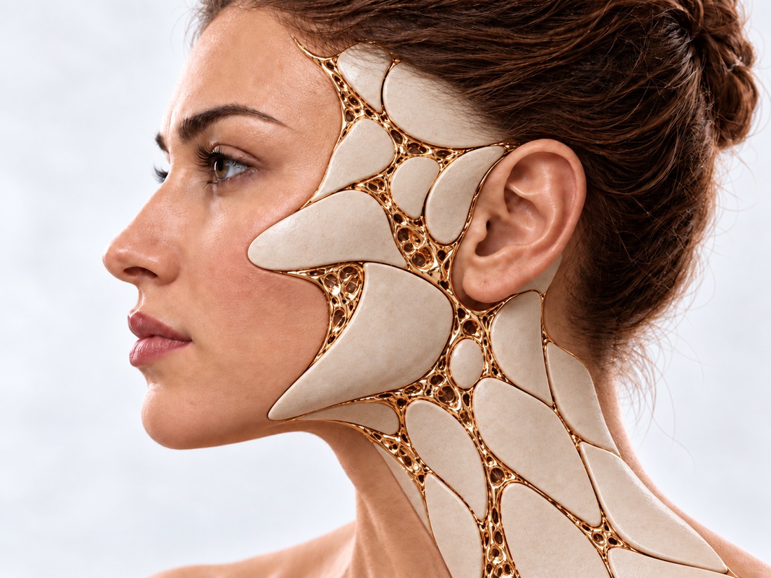
How a
new ear appears.

The gold is marked with the framework from which the ear forms.

01

The frame is made of rib cartilage.

Crunch ribs are cut out and the earshell framework collected cartilage 6 - 8 ribs The ear frame

From the cartilage of several ribs, the surgeon cuts the base and the lunch and sews it into a three-dimensional frame, following the pattern of a healthy ear.

02

Two Stages of Breast Cancer Treatment

The first step is a frame under the skin; the second step is to raise your ears from your head. 1 _ skin frame 2 ears up. It's an interesting wrapper.

After 6-12 months, the ear is removed from the head and a pleat is created on its back. The implant is then inserted under the skin of the temporal area.

03

Implant in the fascia.

The implant is wrapped with viscous-thermal fascia on the receptacles and covered with skin. temporal receptacles EnvironmentTurns the implant. Medpor implant

The artificial framework is wrapped with a thin layer of temporal fascia on its own vessels — it nourishes the skin above the implant.

The schemes are simplified and not scaled up.

When to do
surgery.

  1. Micros: rib cartilage — 6-10 years old

    When the cartilage is sufficient for a framework and the size of the ear for an adult.

  2. Micros: implant — 3-4 years old

    It used to be without a reger, but with the risk of cutting the implant.

  3. After injury or custody

    When the wounds heal and the scars mature, the plan depends on the state of the skin around it.

  4. Interference

    When the swelling drops and the scar stabilizes after surgery.

How the recovery
is going.

The term depends on the method; the plan is individual.

Dressing

First days are protective bandages, skin control over the frame.

Chest

After rib cartilage removal, pain in the scar area for several weeks. Chest deformation is also possible in this area; risk reduces with preservation of ribs.

Ear Protection

For months, they avoid ear pressure and contact sports.

Between Stages

Between the first and second stages, 6-12 months, check-ups.

How much
does it cost.

The value depends on the method, the number of periods and days in the hospital; the surgeon calls it after examination.

From the first
call.

  1. Conversations and materials

    You describe the request and, if you have it, you provide the results of the previous examination and treatment.

  2. Consultation and evaluation

    The doctor determines what situation we are in, what circumstances we need, what we need, and what his limitations are.

  3. Reorganization

    It discusses the next steps, the sequence of phases and the issues of reconstruction.

  4. You don't live in Kiev or abroad?

    Patients come from all over Ukraine and abroad.

What is worth
discussing?

01

How old is the patient, what height and weight?

02

Have they checked the hearing?

03

Was there an ear surgery?

04

Is there a picture of both ears?

Frequently Asked
Questions.

The short answers are that what suits you is what the doctor will tell you.

From what age does the reconstruction of the ear with microthyia?

From his rib cartilage, usually from the age of 6 to 10, when the cartilage is sufficient.

What's better, rib cartilage or implant?

Own cartilage is less likely to be exposed and infected, can grow with the child but requires rib removal (possibly deformation of chest) and two or more stages. Implant – earlier and often in one step, but with risk of piercing and removal.

Will the hearing improve after the reconstruction?

No, reconstruction restores the shape of the ear, hearing evaluates and treats the LOR-licar separately.

Can you raise an ear in a lab?

So far only in clinical research, in normal practice, the ear is modelled from rib cartilage or implant.

When can you fix a bad mistake?

When the edema and the scar stabilizes after surgery, the surgeon's timing is determined on sight.

Sources

13 sources of assessments and studies
ShowTurn to Head
  1. Rib Autograft Versus Porous Polyethylene Implant Outcomes in Microtia Reconstruction: A Meta-Analysis and Systematic ReviewThe Cleft Palate-Craniofacial Journal, 2026doi:10.1177/10556656251349274 ↗
  2. Brent Technique of Repair Versus Nagata Auricular Reconstruction for Microtia Reconstruction: A Systematic Review and Meta-AnalysisJournal of Craniofacial Surgery, 2024doi:10.1097/SCS.0000000000009709 ↗
  3. Autologous Rib Microtia Construction: Nagata TechniqueFacial Plastic Surgery Clinics of North America, 2018doi:10.1016/j.fsc.2017.09.006 ↗
  4. Microtia Ear Reconstruction with Patient-Specific 3D Models-A Segmentation ProtocolJournal of Clinical Medicine, 2022doi:10.3390/jcm11133591 ↗
  5. General and Treatment-Specific Outcomes with Osseointegrated Implants in Auricular, Nasal, and Orbital Prosthetic ReconstructionCraniomaxillofacial Trauma & Reconstruction, 2025doi:10.3390/cmtr18010016 ↗
  6. In Vitro Regeneration of Patient-specific Ear-shaped Cartilage and Its First Clinical Application for Auricular ReconstructionEBioMedicine, 2018doi:10.1016/j.ebiom.2018.01.011 ↗
  7. Posterior Perichondrioadipodermal flap: A Versatile Option for Revision OtoplastyAnnals of Plastic Surgery, 2020doi:10.1097/SAP.0000000000001939 ↗
  8. Complications of Cartilage Sparing Otoplasty: A Systematic Review and Meta-AnalysisJournal of Craniofacial Surgery, 2024doi:10.1097/SCS.0000000000009976 ↗
  9. Revision otoplasty: an algorithmPlastic and Reconstructive Surgery, 2012doi:10.1097/PRS.0b013e31825dc363 ↗
  10. Understanding the unfavorable result after otoplasty: an integrated approach to correctionPlastic and Reconstructive Surgery, 2011doi:10.1097/PRS.0b013e31821e7113 ↗
  11. Complications of otoplasty: a literature reviewJournal of Plastic, Reconstructive & Aesthetic Surgery, 2009doi:10.1016/j.bjps.2008.06.043 ↗
  12. Does postauricular fascial flap reduce suture complications in otoplasty?Journal of Plastic Surgery and Hand Surgery, 2018doi:10.1080/2000656X.2018.1444618 ↗
  13. Updated scar management practical guidelines: non-invasive and invasive measuresJournal of Plastic, Reconstructive & Aesthetic Surgery, 2014doi:10.1016/j.bjps.2014.04.011 ↗

Metaspects and studies on methods described on the page.

VALIKHNOVSKI SURGERY PARTNERS

Your case
deserves attention.

Start consulting to see what questions and materials are needed for the evaluation.

Call Send a request