
FACE / NOSE

Nasal reconstruction.

We restore the shape, framework and breathing function of the nose after trauma, unsuccessful surgery, burns and tumour removal.

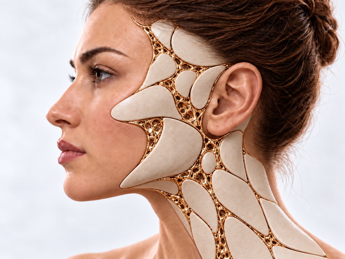
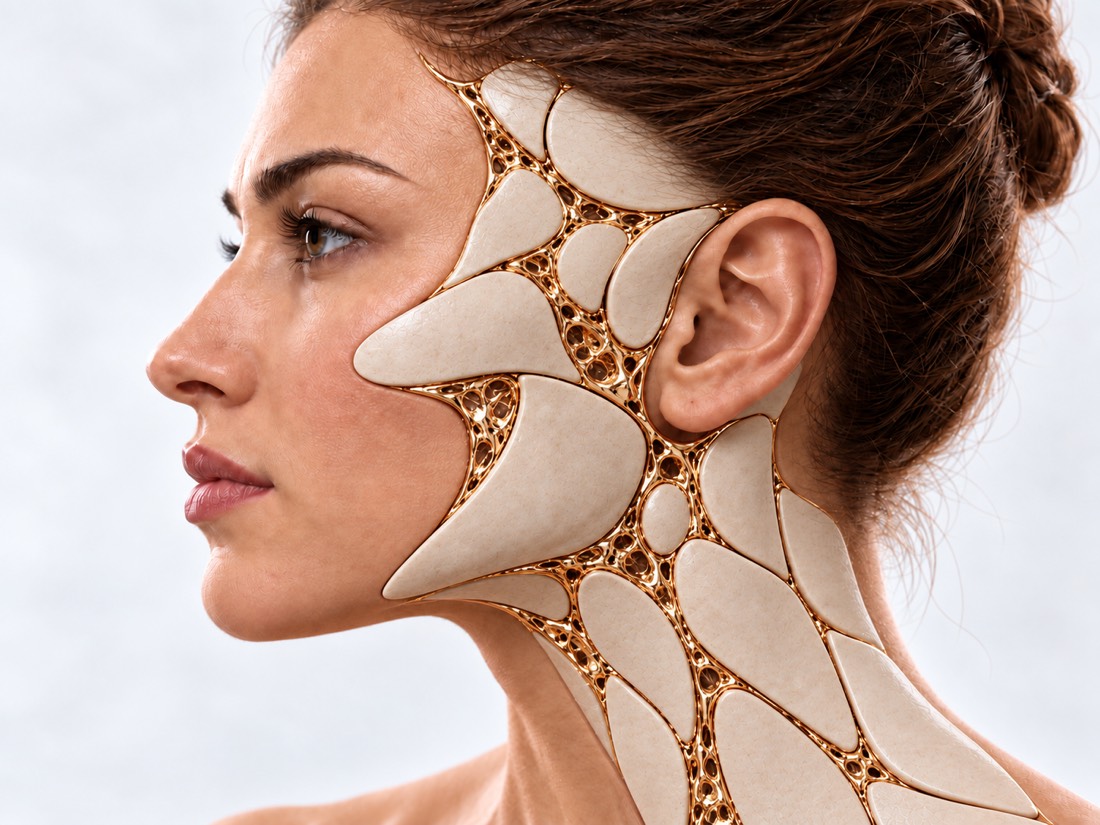
Conceptual visualization directly by "New nose": face with gold lines of reconstructionFIG. 03 / CONCEPTUAL VISUALIZATION

Just injured your nose?

This page deals with routine recovery: first, the doctor examines the injury in the injury or in the emergency room.

The nose changed shape after the impact.

Do not try to straighten it yourself. See a surgeon in the first few days: fresh fractures with dislocation are corrected up to 3 weeks after injury until bone has grown back. Correction does not always restore the nose’s previous appearance – then the shape is later scheduledly correct.

103 or directly to the reception room if:

  • The nose bleed does not stop;
  • Inside the nose is a purple edema that prevents breathing;
  • A transparent watery liquid flows from the nose;
  • You have an eyeache and two eyes.
  • A severe headache with blurry vision;
  • The pain or the truncheon of the neck, or the hands of the hand, shall be punctured;
  • was vomiting, loss of consciousness or broken language;
  • A big wound, or a wound, glass or wreckage.

What is
nose reconstruction.

The nose recommendation is an operation that restores the shape of the nose, its frame, and its breathing.

They are done after injuries and fractures, failed rhinoplasty, septum perforation, burns and tumor removal. Modern reconstruction of the nose and nasal septum in 2026 is based on a three-dimensional, three-component approach. If earlier plastic surgery focused mainly on appearance, today the priority task is to harmoniously combine aesthetics, rigid framework and full breathing.

A surgeon is consulted when the nose has moved or sunk after a stroke, when it looks badly after previous surgery or breathes worse, if there is an opening in the septum or part of the noses is lost.

The specific indications, method and expected result are determined individually by the doctor after consultation and necessary examinations.

From damage
to recovery.

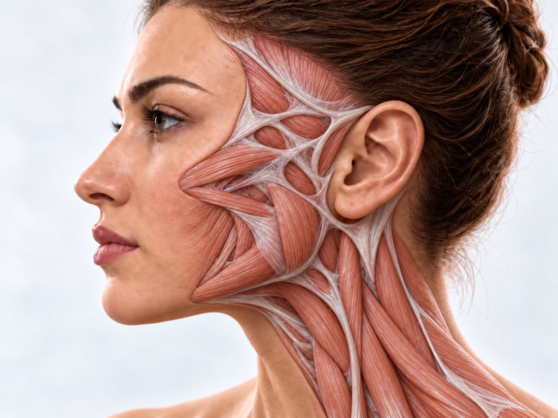
Move the slider. Open anatomic layer means damaged tissue, gold means the reconstruction performed.

The artist is direct, not a picture of the patient.

Golden facial reconstruction in conceptual visualization Opening muscles and facial tissue in conceptual anatomical visualization BACKGROUNDRECONSTRUCTION
FIG. 03.1 / CONCEPTUAL VISUALIZATION, NOT PACENT PHASE

What can be
restored.

Six of the most frequently visited states, each of which has its own page with a detailed answer.

01

Nose after injury

The nose is shifted aside, on the back of the hills, in the west, the partition is distorted, and it's harder to breathe.

How it is treated

Fresh fractures with displacement up to 3 weeks are treated in a closed manner, without any skin incision. If the bone has already grown wrongly, the nose is aligned according to plan, usually after 2-6 months: as indicated, bones are cut (osteotomia) and repositioned, cartilage straightened.

Nasal deformation after injury
02

Saddle-shaped nose.

The back's jammed because the partition's cartilage is broken or lost.

How it is treated

The subluxation occurs after an injury, hematoma or previous surgery; other causes are certain diseases (granuloma with polyangiitis, recurrent polychondritis) and cocaine use. They are excluded before surgery: in the case of disease, only operated on a stable remission.

Saddle nose
03

Failed Rhinoplasty

Asymmetry, the rest of the hump, back impact, deformed tip, worse breath.

How it is treated

Repeated operations are more complicated than primary ones: tissues changed by scarring, the cartilage of the septum may be missing. The surgeon restores the framework of your nose – straightens and strengthens the cartilagine, if necessary taking cartilages from an ear or rib.

Nasal deformation after phinoplastics
04

Curved septum.

One or both half of the nose is breathing badly, often after injury or prior surgery.

How it is treated

The septum is leveled (septoplasty): cartilage and bone are straightened, preserving the mucous membrane. If there is no cartilage, fragments are collected on a thin plate that is then sucked out. For details see section "Methods".

Swaller of noses
05

Perforation of the septum.

Scratch hole in the partition: breath whistle, pick, bleeding, bookmark.

How it is treated

Not every perforation requires surgery. If there are no symptoms, observation may be sufficient; for symptomatic cases, moisturising treatment and sometimes a silicone obturator can help. Before surgery, the cause is established. The opening is closed with the patient’s own tissue, using mucosal flaps and, when needed, an interposition graft. Larger perforations are more difficult to close.

Nasal septal perforation
06

Loss of part of the nose.

After custody, tumour removal, bite or severe injury, there is a lack of skin, cartilage or all of the nose.

How it is treated

The nose is restored in three layers: the inner ridge, cartilage frame and skin. For the skin most often a forehead bone is taken. Reconstruction is done in several stages.

Recommendation for burns

How do they restore
the nose.

Modern techniques are divided into anatomical components of the nose that require repair.

Core procedures

They start most interventions after trauma and failed surgery.

OperationWhen it is suitable
Screaming exercisefresh fracture with displacement, up to three weeks after injury
OsteotomyThe nose bone has grown wrong: it is scattered and moved.
SeptorplasticsYou need to restore both the shape of the nose and the partition for one operation.
Replanting ballsIncendiary back, weak frame, repeat operations; cartilage — partition, ears or ribs
Closure of perforationhole in the barrier with complaints: mucous, if necessary with interlayer
01

Reconstruction of the nasal septum (septoplasty and closure of perforations)

The septum is the main "support" of the nose and regulator of air flow. Modern approaches allow it to be restored even in conditions of massive destruction (after injuries, burns, necrosis or unsuccessful operations).

Low injury Endoscopy Septoplastics
The surgeon operates under the control of an endoscope — a thin optical tube with a camera — through an incision inside the nose: he can see better the area of curvature and aligns cartilage and bone, preserving the mucous membrane.
Biodegradable PDS (polyoxanone)
In severe cartilage deficiency, the crushed fragments of the membrane are assembled and fixed on a thin synthetic PDS plate. The plate supports the fragment as they grow and completely resorbs in several months.
Closure of perforations with an acellular dermal matrix (ADM)
A cellless dermal allograft (ADM) is placed between the mucous membrane flaps. The matrix serves as a framework on which its own mucosa grows.
Silicon splints with air ducts
Instead of tampons, anatomical silicone tires (splint) with internal airways can be installed after surgery, which allows partial nasal breathing already after the operation.
02

Total and subtotal reconstruction of the nose.

In the case of a complete or partial loss of the nose (for example, after a burn or oncoresection), surgeons restore it using a three-layer principle.

A. Internal lining: radial forearm free flap
Without restoring the inner layer, nasal passages become narrowed and the nose tightens and scars. A skin flap with vessels is taken from the forearm. Its vessel is sutured under a microscope to facial blood vessel, and then the flaps are twisted into the noses to create a live internal fold.
Carcass: autotransplantation of rib or ear cartilage
The main material remains the patient's own cartilage. Modern surgeons use a method of crushed cartilages in special shells (like "sausages") for delicate restoration of the back of your nose, or they cut out integral L-shaped beams from ribs that are fixed with titanium micro screws if necessary.
B. 3D modelling and navigation
Before surgery, an accurate 3D model of the future nose is created using a CT scan. During the procedure, the surgeon compares every millimeter of cartilage structure with a computer template.
C. Skin: paramedian forehead flap
The skin is cut off on the forehead (leaving it attached to the vascular leg near the eyebrow) and moved onto the nose. The foreheart skin matches well in color and texture. Recent modifications of the design allow for its transplantation with minimal damage to hair growth line.
Previous expansionary dermotension
If there is not enough skin on the forehead (due to concomitant burn scars), a silicone balloon is pre-inserted underneath it for gradual stretching in order to obtain the required volume of healthy tissue.
03

Revision surgery and tissue-preserving techniques

Some of the calls are the effects of previous nose surgery, and it's important to keep your tissue where possible.

Secondary and third-line phinoplastics
The surgeon assesses what has changed under the skin since previous surgery and restores the nose frame: straightens and strengthens cartilage, if necessary taking own cartilages from ear or rib.
Five instruments
They are used to perform osteotomies and model the bony part of the back. The piezo is an instrument, not a separate type of surgery: it is up to the surgeon whether or not he needs one in each case.
Principles of Teorhinolasty Method
The original method of conservative rhinoplasty, developed primarily for primary operations: the cartilage, bones and ligaments of the nose are mostly displaced and only minimally excised, tissue is delaminated under the ridge. In repeated and reconstructive surgeries, the clinic applies its principles where anatomy allows it.

How to correct
a failed rhinoplasty.

Repeat surgery is rare after the first operation, more frequent after secondary: 3.1% vs. 11.0% (175,842 patients following septoplasty in three US states, at least 2 years of follow-up).

01

Why do they operate again?

Breathing and asymmetrics
Learn more

Among the 100 patients who had a second rhinoplasty, difficult breathing (65%), back asymmetry (33%) and nostril alignment (18%) were among the most common complaints. Almost two-thirds of those reviewed reported both shape and breath problems, so the plan for reoperation takes into account both.

Mean of nose
Learn more

Collapse of the inner nasal valve, back deformation into an “inverted V” and asymmetry of middle third are among the most common reasons for re-referral. The main tool here is spreader grafts: cartilage strips along the septum that widen and level out the middle part of your nose.

02

How to restore the framework?

Rib cartilage
Learn more

When there is no more cartilage in the septum, cartilages are taken from an ear or rib. For rib cartillary grafts, a meta-analysis of 20 studies (1648 patients) showed that graft curvature occurred in 3.05%, resorption in1.2%, infection in2.45% and irregularity in3,53%; repeated surgery was required in7.

“Parrot beak” deformity
Learn more

Excess tissue above the tip of the nose after rhinoplasty – soft tissue or cartilage. For the soft-tissue variant, several methods have been described to prevent and treat it without surgery, but their effectiveness has not yet been proven by comparative studies.

03

What to expect from the revision?

More complex operation
Learn more

In repeated surgeries, complex reconstructive techniques and grafts taken outside the nose were more commonly used. Another surgery after revision was also needed more often: 23.9% compared to 10.5% after primary (245 patients from one specialized clinic).

What happens
during reconstruction.

Three diagrams in the "Methods" section. Golds are marked with tissue that restores the defect.

01

Three layers of the nose.

Three layers of nose structure: skin, cartilage and interior rack person Skin ♪ Browsing ♪ B· framework Brisket or ears A · scrubbing Mucosa or tissue flap

The nose has three layers: skin on the outside, cartilage and bone framework inside and mucous membrane inside. When a part of the nose is lost, each missing layer is rebuilt: without the bridge and frame, the skin tightens with scarring and the noses lose shape.

02

Forehead flap

Paramedian forehead flap: designed on the forehead and transferred to the nose in two stages Klapot 1 · Transport On your forehead. 2 legs cut off.

The flap is cut off on the forehead and returned to the nose, leaving a nourishing leg near the eyebrow. When the flaps have survived in their new place, the leg is then cut out at the next stage and the front is sewn together.

03

Closure of perforation.

Cross-section of a septal perforation and closure with mucosal flaps and an interposition graft openingMist · cartilage · mucous 1 air whistles Layercartilage, fascia Klapty mucous 2 holes closed

The opening is closed with flaps of the own mucosa on both sides, and between them a layer of cartilage, fascia or biomatrix is placed if necessary.

The schemes are simplified and not scaled up.

When to do
surgery.

  1. Fresh fracture: up to 3 weeks

    Until the bones have grown, they are exercised in a closed manner without any incisions. The hematoma of the septum is opened as soon as possible, otherwise it can destroy cartilage.

  2. Long-term distortion: 3-6 months from now

    When the swelling stabilizes, the nose is evenly aligned.

  3. Repeated Rinoplastics: Not before a year later

    About this time the final shape of the nose is formed after previous surgery. Earlier — if breathing is severely impaired or there are complications.

  4. Nose part loss: in stages

    After tumour removal, reconstruction is planned once complete excision has been confirmed. Reconstruction with a forehead flap usually takes two or three stages, several weeks apart.

How the recovery
is going.

Terms depend on the number of transactions; the exact plan is individual.

First weeks of pregnancy

On the back of the nose, the home country, and the seat, it's harder to breathe with the nose, and the synetus and the main swelling take weeks.

Up to one year

The final form of the nose, especially the tip, is about a year long and sometimes longer after repeated operations.

What to avoid

Do not cut yourself or wear glasses on the back of your nose until permission is given by the surgeon. Sports and exercises are allowed after permissions have been obtained.

When to call 103?

Bleeding does not stop despite squeezing; shortness of breath or chest pain; high fever with a rash, vomiting or dizziness, especially with swabs or tires in the nose. Temperature 38 °C and above or pus — same day to surgeon.

How much
does it cost.

The value of the operation is called after examination, depending on:

  • Original or repeated operation;
  • Is there a need for an ear or a rib cartilage?
  • Refurbish the partition;
  • Number of periods and days in hospital.

From the first
call.

  1. Conversations and materials

    You describe the request and, if you have it, you provide the results of the previous examination and treatment.

  2. Consultation and evaluation

    The doctor determines what situation we are in, what circumstances we need, what we need, and what his limitations are.

  3. Reorganization

    It discusses the next steps, the sequence of phases and the issues of reconstruction.

  4. You don't live in Kiev or abroad?

    The clinic is visited by patients from all over Ukraine and other countries. Send photos and extracts – the doctor will pre-evaluate your case remotely and tell you what to bring for consultation.

What is worth
discussing?

01

What happened to the nose and when?

02

Is it difficult to breathe with one or both noses?

03

What kind of nose surgery have you had?

04

Is there a photo before the injury or surgery, a discharge, C.T.?

05

What chronic diseases and medications?

06

What's more important to you than breathing or looking?

Frequently Asked
Questions.

The short answers are that what suits you is what the doctor will tell you.

When can I do a rerun of Rinoplastics?

Usually not earlier than a year after the previous operation: about that much is formed the final shape of the nose. Earlier - if there are serious breathing disorders or complications.

Can you level your nose after a fracture without incisions?

Yes, if the fracture is fresh: up to 3 weeks after injury bones are closed. Treatment does not always restore their previous appearance. If the bone has already grown incorrectly, nose straightening is planned, usually after 2-6 months.

Where do they get the cartilage for the nose reconstruction?

If it is missing, for example after previous operations, from the eardrum or rib. The choice depends on how much cartilage is needed and for which part of the nose.

Can a septal perforation close on its own?

Usually not. But not every one requires surgery: if there are no complaints, the hole can only be observed. Whether to operate or not, a surgeon decides after examination and finding out the cause.

Can the nose be restored if part is lost?

Yes. Every missing layer is restored: the lining, cartilage frame and skin – most often a flap from the forehead. This reconstruction takes several stages.

Will there be tampons in the nose after the surgery?

Instead of gauze pads, soft silicone tires with air ducts are often used, through which you can partially breathe through your nose. What exactly will happen to you, the surgeon will tell before surgery.

How long does the swelling last after the nose surgery?

Bruising and basic swelling go away in a few weeks, but the final shape of the nose, especially at the tip, takes about one year. After repeated operations, swelled areas may last longer.

Sources

14 NHS, StatPearls and peer-reviewed surveys
ShowTurn to Head
  1. Broken noseNHS (United Kingdom)nhs.uk/conditions/broken-nose ↗
  2. Advancements in the Application of Artificial Materials in Nasal SurgeryJournal of Craniofacial Surgery, 2025doi:10.1097/SCS.0000000000011285 ↗
  3. A Surgical Strategy for Three-Layer Structure Reconstruction in Total Nasal DefectJournal of Clinical Medicine, 2026doi:10.3390/jcm15145459 ↗
  4. Designing a Nasal Lining-Framework Complex for Reconstructing Total Nasal DefectsOtolaryngology–Head and Neck Surgery, 2024doi:10.1002/ohn.1006 ↗
  5. Advances in forehead flap nasal reconstruction: from design to divisionCurrent Opinion in Otolaryngology & Head and Neck Surgery, 2026doi:10.1097/MOO.0000000000001134 ↗
  6. Surgical repair of large nasal septal perforation: a systematic review and meta-analysisEuropean Archives of Oto-Rhino-Laryngology, 2026doi:10.1007/s00405-025-09913-9 ↗
  7. Septal PerforationStatPearls, NCBI Bookshelfncbi.nlm.nih.gov/books/NBK537208 ↗
  8. A New Understanding and a Minimalist Approach for RhinoplastyPlastic and Reconstructive Surgery, 2023doi:10.1097/PRS.0000000000010213 ↗
  9. Revision Rates and Risk Factors of 175 842 Patients Undergoing SeptorhinoplastyJAMA Facial Plastic Surgery, 2016doi:10.1001/jamafacial.2015.2194 ↗
  10. Frequency of the preoperative flaws and commonly required maneuvers to correct them: a guide to reducing the revision rhinoplasty ratePlastic and Reconstructive Surgery, 2013doi:10.1097/PRS.0b013e3182a01457 ↗
  11. Differences between Primary and Revision Rhinoplasty: Indications, Techniques, Grafts, and OutcomesPlastic and Reconstructive Surgery, 2021doi:10.1097/PRS.0000000000008216 ↗
  12. Spreader Grafts in Functional RhinoplastyFacial Plastic Surgery, 2016doi:10.1055/s-0035-1570127 ↗
  13. Complications Associated with Autologous Costal Cartilage Used in Rhinoplasty: An Updated Meta-AnalysisAesthetic Plastic Surgery, 2023doi:10.1007/s00266-022-03075-3 ↗
  14. Prevention and non-surgical treatment of soft tissue polly beak deformity after rhinoplasty: a scoping reviewJournal of Cranio-Maxillofacial Surgery, 2023doi:10.1016/j.jcms.2023.02.002 ↗

The recent injury is recommended by NHS, while the rest are peer-reviewed surveys of methods described on the page.

VALIKHNOVSKI SURGERY PARTNERS

Your case
deserves attention.

Start consulting to see what questions and materials are needed for the evaluation.

Call Send a request