We remove sarcomas and other soft-tissue tumours while aiming to preserve the limb and its function, reconstructing tissue, vessels and nerves during the same operation when needed.
Most of the seals in the soft tissue are good, but some of the signs are the reason to see a doctor as soon as possible.
To the doctor without delay if the thickening:
grows or becomes more than 5 cm;
lies deep under the muscle or fascia;
It hurts;
appeared again at the disposal site.
Don't delete "just like that".
If sarcoma is suspected, MRI and biopsy are performed first at the site of surgery. Removal without examination can complicate further treatment.
01 / WHAT IT IS
What is soft tissue oncology.
Soft tissue oncology is the removal of sarcomas and other tumors from muscle, adipose fiber and connective tissue with restoration of what had to be removed.
Modern technologies of surgical treatment of soft tissue tumors (especially sarcoma) are based on the principle of maximum preservation of organ and limb function without compromising oncological radicality. Thanks to the combination of surgery with radiation therapy, today about 90% of patients with limber sarcomas undergo organ-preserving operations, avoiding amputationes.
They are consulted with a dense growth, confirmed sarcoma, recurrence after previous surgery or tissue defects following tumor removal.
Treatment of sarcoma is planned together with an oncologist and a radiation therapist. This is a general description of the direction: indications and method are determined by the doctor after examinations.
02 / With PM, it's dead.
When you need a surgeon-oncologist.
Six situations that are most common.
01
A dense, growing growth.
The nest under the skin or in the muscle increases, pains, or lies deep.
How it is treated
First the MRI, then the biopsy, the fat needle with histology, only after the diagnosis, is there an operation planned.
The affected artery, and if necessary the vein, is removed along with the tumor and replaced by a prosthesis or an own veins; the nerve is plasticized where possible.
Basic surgical techniques and approaches that shape modern soft tissue oncology.
01
Preoperative tumor reduction.
To make surgery possible or less traumatic, it may be preceded by preoperative treatment – radiation or chemotherapy prescribed by an oncologist.
Isolated hyperthermic perfuse of limbs (ILP)
A procedure for patients with large tumors of the arms or legs. Surgeons temporarily isolate the limb’s vascular canal from the general bloodstream using a circulation device and wash it with a heated solution of high-dose drugs (tumor necrosis factor alfa (TNF-α) and melphalan). This often shrinks the tumour, after which it can be removed while preserving the extremity.
02
Digital Pregnancy Planning
If necessary, the MRI and CT data are combined into a three-dimensional model prior to intervention.
3D modelling
The MRI and CT data are combined into a three-dimensional model of the tumor. The surgeon sees how the neoplasm is located relative to arterial vessels, nerve trunks and bones.
Added reality (AR)
During surgery on the surgeon's screen or glasses, a 3D model is used to guide within the resection.
03
Block deletion (en bloc)
The main standard for sarcoma removal is a wide anatomical resection in one block. The tumor is removed together with the pseudocapsule and layer of surrounding healthy tissues.
Control of the cutting edge (R0)
The surgeon’s goal is to obtain microscopically clear margins around the resected specimen, with no tumour cells at the cut edge. A pathologist makes the final margin assessment; when necessary, selected margins are examined during surgery.
04
Microsurgical and vascular reconstruction.
When radical excision requires the removal of important structures, reconstruction is performed at the same time as tumor removal.
Autologous transfer of a musculocutaneous flap
Large defects are closed with the patient's own flaps on the vascular leg or microvascular anastomosis (for example, from the widest muscle of the back or anterior surface of thigh).
Protection of blood vessels
If the sarcoma has grown into a large artery or vein, the affected area is removed and replaced with synthetic prostheses or an own veins, restoring blood supply to the limb.
Nerve plastic
In case of forced removal of the motor nerves, plastic surgery (autoneuroplasty) is performed which can partially restore mobility in the limb in future.
05
Intraoperative radiation therapy (IORT) and brachytherapy
IORT
During surgery, after the removal of sarcomas, surgeons and radiation therapists use mobile accelerators to irradiate the tumor bed with a single high dose.
Brachytherapy
Catheters are placed in the tumor bed, the wound is sutured and then a radioactive source is administered through them precisely to the area of highest risk for recurrence.
04 / HOW IT GOES
What happens during the operation.
The gold marks the tissue that the defects restore.
01
Single block removal.
The tumor is removed not by "peeling", but with the surrounding layer of healthy tissue, without revealing it. A pathomorphologist checks for tumor cells along the edges.
02
Vascular prosthesis.
If the tumor has grown into an artery or vein, this area is removed along with the tumour and blood flow is immediately restored by a synthetic prosthesis or the patient's own veins.
03
Closing the defect with a flap.
If the wound cannot be simply sutured, it is covered with a flap – skin with muscle or fat on its own vessels – often in the same operation.
The schemes are simplified and not scaled up.
05 / PHASE
How is the treatment going.
MRI and biopsy
Diagnosis is behind histology; biopsy is desirable where they operate.
Multidisciplinary team review
The surgeon, the oncologist, and the sales therapist decide if they need treatment before surgery.
Operation and reconstruction
The removal of a single block and the closure of a defect are more often than not the same operation.
Histology and oversight
After evaluation of the margins, radiation or chemotherapy as indicated; regular MRI of surgery site and CT or chest X-ray.
06 / AFTER OPERATION
How the recovery is going.
Terms depend on size; the plan is individual.
Hospital stay
After major resections and flap reconstruction, several days of inpatient monitoring are usually required.
Healing
After preoperative radiation therapy, healing complications occur more often; to close the wound with well-blown tissues, a surgeon may use a flap.
Rehabilitation
After the muscles and nerves are removed, the exercise is to restore strength and movement.
Follow-up
Regular examinations, MRI and CT or chest X-rays for several years.
07 / AFTER OPERATION
Complications, rehabilitation and supervision.
After sarcoma is removed, three things are important: healing the wound, returning the function, and regular monitoring.
01
Wound Healing
How Often Complications Can Be
In a meta-analysis of 21 studies on limb sarcoma (5,628 patients), wound complications had a 30.2% risk and reoperation was required in 13.4%. The risk is higher for leg tumors, diabetes, smoking, obesity and radiation.
Pre- or post-op radiation
In a randomized study, preoperative radiation was twice as likely to cause wound complications (35% vs. 17%) than non-radiotherapy. The studies differ on the impact of preoperational irradiation on survival. Therefore, treatment is chosen based on tumor size and location, with an oncologist, radiotherapist and surgeon working together to develop a plan.
Why put it down?
A systematic review associates flap closure with fewer complications, especially when radiation is planned after surgery; it is recommended to be performed in the first 3-6 weeks following reconstruction. The limb was preserved in more than 90% of cases for all types of reconstructions and joint planning by cancer surgeon and plastic surgeons is associated with less wound complication.
02
Restoration of function.
Rehabilitation from the beginning
After treatment, movement restrictions, pain, swelling of the limb and psychological difficulties are possible. Rehabilitation is advised to be planned individually and started early – ideally before the first stage of treatment.
Limphedema
In one cohort of 289 patients, lymphoedema (persistent swelling of the limb) appeared in 10.5% and was severe in about 7%. Lymphedema occurred more frequently with tumors larger than 5 cm and deeper.
03
Follow-up after treatment
Regular inspections
Even after optimal treatment, sarcoma can recur and follow-up is necessary: at least regular checkups and chest x-rays. The frequency and scope of the screening are determined by the risk of relapse.
08 / INSTALMENT
How much does it cost.
The cost depends on the size and location of the tumor, the extent of reconstruction and days in hospital; it is appointed by the surgeon after examinations.
09 / PACIENT SHOTS
From the first call.
Conversations and materials
You describe the request and, if you have it, you provide the results of the previous examination and treatment.
Consultation and evaluation
The doctor determines what situation we are in, what circumstances we need, what we need, and what his limitations are.
Reorganization
It discusses the next steps, the sequence of phases and the issues of reconstruction.
You don't live in Kiev or abroad?
Patients come from all over Ukraine and abroad.
10 / For Consultation
What is worth discussing?
01
When did the sealing start, and how did it change?
02
Is this MRI, CT, biopsy, histology?
03
Have these sites been operated or irradiated?
04
What chronic diseases and medications?
11 / FOOD AND REPLIES
Frequently Asked Questions.
The short answers are that what suits you is what the doctor will tell you.
Can you keep your limb in sarcoma?
In most cases, this is the case: about 90% of patients with limb sarcomas are operated on and their limbes are preserved. Amputation is necessary when otherwise the tumor cannot be completely removed.
Can you just cut the seal?
If it is a sarcoma, the "excision" without a reserve of healthy tissue increases the risk of recurrence and then repeated, larger surgery is needed. First — MRI and biopsy.
If the tumor was removed, why radiation therapy?
In order to reduce the risk of a repeat tumor in the same location, it is assigned before or after a constitutional operation.
Do they do the reconstruction at once?
If the wound cannot be simply sutured, it is usually covered with a flap in the same operation so that the injury heals and further treatment can begin on time.
How often is post-op examination required?
Regularly over several years: MRI of the surgical site and CT or chest x-ray, because sarcomas metastase more often to the lungs. The schedule is determined by the oncologist.
Soft tissue and visceral sarcomas: ESMO-EURACAN-GENTURIS Clinical Practice Guidelines for diagnosis, treatment and follow-upAnnals of Oncology, 2021doi:10.1016/j.annonc.2021.07.006 ↗
Role of isolated limb perfusion with recombinant human tumor necrosis factor α and melphalan in locally advanced extremity soft tissue sarcomaCancer, 2016doi:10.1002/cncr.29991 ↗
Intraoperative radiation therapy (IORT) for soft tissue sarcoma - ESTRO IORT Task Force/ACROP recommendationsRadiotherapy and Oncology, 2020doi:10.1016/j.radonc.2020.07.019 ↗
Preoperative versus postoperative radiotherapy in soft-tissue sarcoma of the limbs: a randomised trialThe Lancet, 2002doi:10.1016/S0140-6736(02)09292-9 ↗
Lower Extremity Reconstruction After Soft Tissue Sarcoma ResectionClinics in Plastic Surgery, 2021doi:10.1016/j.cps.2021.01.007 ↗
Amputation in patients with extremity soft tissue sarcoma: the experience of an East Asian referral centerBMC Cancer, 2024doi:10.1186/s12885-023-11813-2 ↗
Late Local Recurrence and Metastasis in Soft Tissue Sarcoma of the Extremities and Trunk Wall: Better Outcome After Treatment of Late Events Compared with EarlyAnnals of Surgical Oncology, 2021doi:10.1245/s10434-021-09942-8 ↗
Preoperative versus postoperative radiotherapy in soft-tissue sarcoma of the limbs: a randomised trialThe Lancet, 2002doi:10.1016/S0140-6736(02)09292-9 ↗
Risk factors for postoperative wound complications after extremity soft tissue sarcoma resection: A systematic review and meta-analysesJournal of Plastic, Reconstructive & Aesthetic Surgery, 2019doi:10.1016/j.bjps.2019.05.041 ↗
Therapeutic options and postoperative wound complications after extremity soft tissue sarcoma resection and postoperative external beam radiotherapyInternational Wound Journal, 2018doi:10.1111/iwj.12851 ↗
Soft tissue reconstruction techniques after musculoskeletal tumor resection: A systematic review of gastrocnemius flap, free flap, local flap, and orthoplastic reconstructionEuropean Journal of Surgical Oncology, 2026doi:10.1016/j.ejso.2026.112077 ↗
 Discuss your case
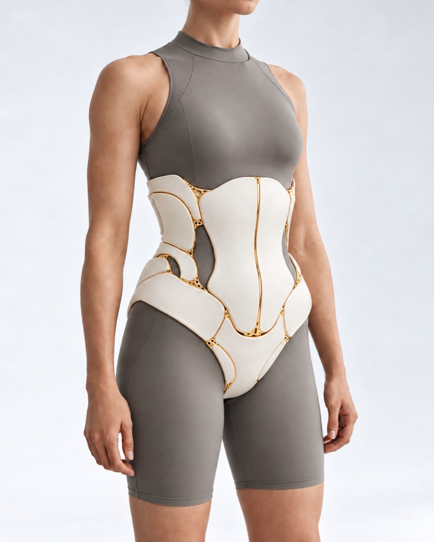 FIG. 11 / CONCEPTUAL VISUALIZATION