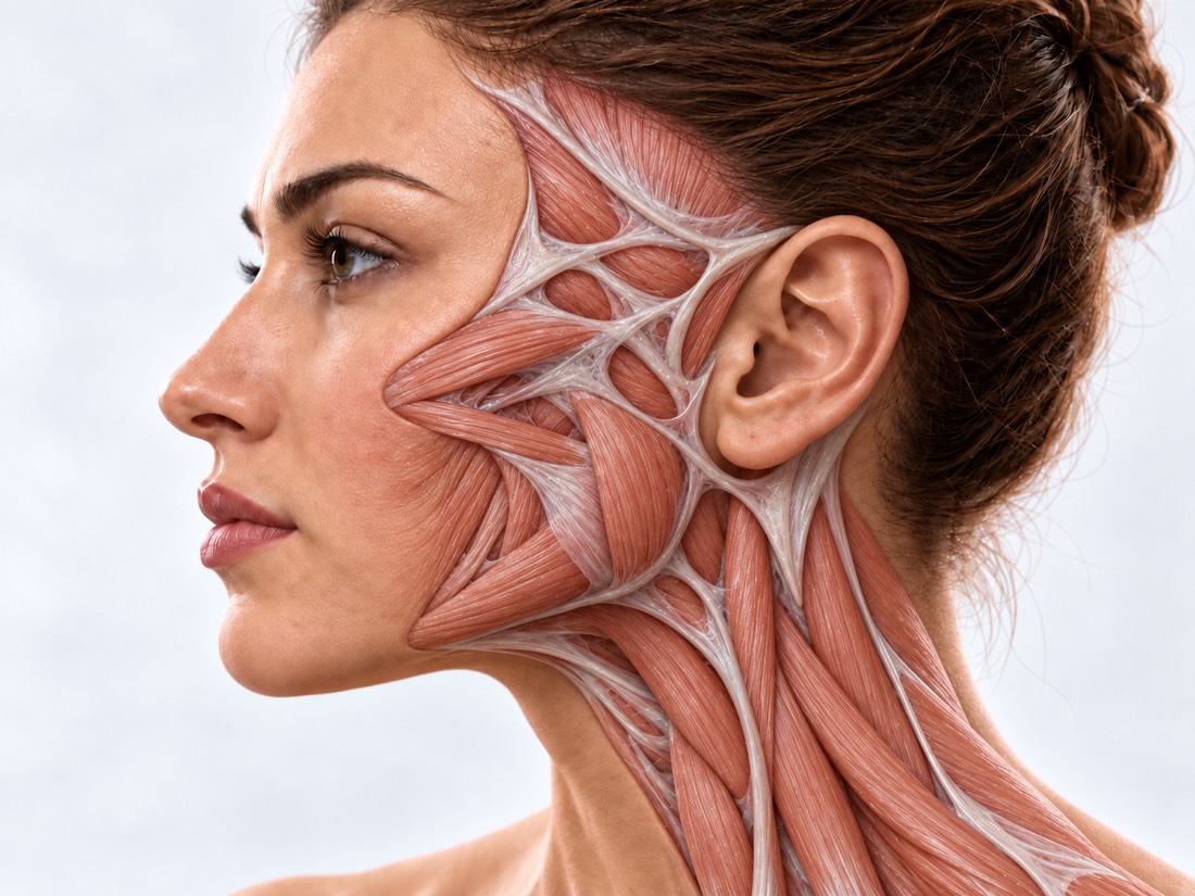
CONDITION / FACE

Basal cell carcinoma of the face and head.

Excision of a basal cell carcinoma is the main treatment for the most common skin cancer: the tumor is excised with a margin of healthy tissue and the edges are examined under a microscope. If the edges are clean, the defect on the face can be closed in the same operation.

Conceptual visualization: restoration of facial skin after tumor removalCONCEPTUAL VISUALIZATION
What it is
Malignant tumor from the cells of the upper layer of the skin; the most common skin cancer.
Where
Most often, on skin exposed to the sun: on the head and neck - 40–64% of basal cell carcinomas.
What is dangerous?
It rarely spreads to other organs, but without treatment it can grow deep.
Who treats it
The surgeon removes and restores the skin; drugs and radiation are prescribed by an oncologist if necessary.

What is
basal cell carcinoma

Basal cell carcinoma, or basal cell carcinoma, is a malignant tumor that begins in the upper layer of the skin. It is the most common skin cancer in fair-skinned people, and it is becoming more common.

Basal cell carcinoma rarely spreads to other organs, so additional examinations are needed only when it is very large. But without treatment, the tumor can grow and grow deeper — into cartilage, muscles, nerves, or bone.

If basal cell carcinoma is detected early, it is usually curable. On the face, two things are important at the same time: remove the tumor completely and preserve the appearance and functions of the eyelids, nose, and lips. Therefore, removal and plastic surgery of the defect are planned together.

This is general information. The doctor will tell you what will suit you after the examination.

How does it look
basal cell carcinoma

Basal cell carcinoma appears more often on the skin exposed to the sun: on the face, ears, scalp. It can look different - like a nodule, a spot or a wound.

01

A shiny knot

Pink smooth rise with clear edges.

02

A flat spot

Pink or red spot; it may be darker on dark skin.

03

A wound that does not heal

The area bleeds, crusts or scabs and does not heal for more than 4 weeks.

04

Changes

The formation grows, changes color or surface, itches or hurts.

05

Near the eye

Basal cell carcinoma is the most common malignant tumor of the eyelids. Nearby there are structures important to the eye, so the removal and plastic surgery are planned especially carefully here.

Neoplasms of the eyelids

Why does it occur?
basal cell carcinoma

The main reason is ultraviolet. The rest of the factors show who should pay more attention to their skin.

Sun and solarium

Ultraviolet light is the main cause of basal cell carcinoma.

Skin type

Light skin that burns easily is the main risk factor.

Age and gender

Basal cell carcinoma occurs more often in the elderly and in men.

Precancer and skin cancer in the past

Actinic keratosis or already removed skin cancer increase the risk of new tumors.

Precancerous skin diseases

Depressed immunity

In particular, after an organ transplant, when a person takes drugs that suppress the immune system.

When to seek medical advice?

Basal cell carcinoma is a planned situation: it usually does not require emergency care. But you shouldn't delay: the earlier skin cancer is detected, the easier it is to treat. 103 may be needed only because of wound complications after surgery.

103 immediately if:

  • bleeding from the wound does not stop, even if it is pressed;
  • the blood gushes, bright red, hard to stop.

To the doctor:

On the same day: to the surgeon who operated, if the wound after removal is swollen, red, hurts more and more, pus flows from it, or you feel bad and have a fever. Scheduled, without delay: the formation on the skin grows, has changed color or surface; the area hurts, itches, bleeds or crusts for more than 4 weeks. A new birthmark or a change in an old one can be a sign of melanoma - it is a more dangerous skin cancer, so show it to a dermatologist or oncologist without delay; if the doctor suspects melanoma, the biopsy and the oncologist's consultation are performed urgently, without waiting for an appointment.

How we make the
diagnosis

  1. Examination and dermatoscopy

    The doctor examines the formation through a special magnifying glass; by the characteristic signs of a basal cell carcinoma, it is often recognized already on examination.

  2. Biopsy

    If there is doubt, a piece or the entire formation is sent to the laboratory. The result is usually ready in 2-4 weeks.

  3. Risk assessment

    Size, margins, type of tumor under the microscope and location: on the face, the risk of recurrence is higher and the tissue supply for plastic surgery is smaller.

  4. CT scan or MRI if needed

    If there is a suspicion that the tumor has grown along the nerve or into the bone, or the basal cell carcinoma is very large.

How we treat
basal cell carcinoma

According to European guidelines, the treatment of first choice is complete removal of the tumor. Other methods are reserved for low-risk superficial basal cell carcinomas or for those who cannot be operated on.

01

Removal

The goal is to remove the tumor completely.

Stock of healthy tissues
According to the British guidelines, a basalioma of up to 2 cm of non-aggressive type is removed, retreating 4–5 mm. In a meta-analysis of 16,066 tumors, with a margin of 5 mm, it returned in 0.39% of cases, with a margin of 2 mm — in 3.96%.
Edge control
The removed tissue is examined under a microscope. If tumor cells remain on the edge, the basal cell carcinoma returns on average in 27% of cases, so such an edge is removed again.
Inspection during operation
The edges can be examined urgently while the patient is still in the operating room. In a study of 1,082 basal cell carcinomas of the face, this analysis coincided with the final one in 98.8% of cases; primary basal cell carcinoma returned in 1.6%.
Operation Moss
The tumor is removed in layers and the entire edge is examined each time. The instruction advises it for high-risk basal cell carcinomas, recurrent and on critical areas of the face.
02

Closing the defect

Immediately or after histology.

Immediately
If the edges are clean, the wound is closed in the same operation: sutured, local flap moved, or skin grafted. Large defects are closed with more complex flaps.
Later on
If the risk of recurrence is high, plastic surgery is postponed until histology confirms clear margins. In a study of 633 defects, delayed repair did not increase the risk of infection or flap death.
Risks
Wound infection, bleeding, graft death are possible. There are more complications in smokers, with larger and through defects.
03

Without surgery

When surgery is not possible or necessary.

Creams and photodynamic therapy
For low-risk superficial basal cell carcinomas. They are less effective than surgery—after three years, basal cell carcinoma returned in 16.4% versus 1.6% after excision—but the appearance of the skin after them is usually better.
Radiation therapy
An alternative for those who cannot have surgery or who refuse it, especially in old age. It is prescribed and conducted by a radiotherapist.
04

Basal cell carcinoma has started

Treatment is conducted by an oncologist.

Consilium
Basal cell tumors, which are difficult to treat, are recommended to be discussed at a consultation of specialists.
Medicines
If the tumor cannot be removed or has spread, the oncologist may prescribe drugs that block the tumor's growth signals or immunotherapy.
05

After treatment

New basal cell tumors
The risk of a new tumor is high: according to a meta-analysis, within three years — 44%. Therefore, it is recommended to examine the skin at least once a year.
Late relapses
In a randomized trial of facial basal cell carcinoma, 56% of primary tumor recurrences occurred after 5 years, so follow-up is long after high-risk basal cell carcinoma.
Sun protection
Sunscreen is a proven way to prevent skin cancer.

How
we treat it.

Basal cell carcinoma of the face and head is treated in the "New Face" direction at Dr. Valikhnovskyi's Institute of Surgery: the clinic removes the tumor with edge control and restores the defect, often in the same operation. If the basal cell carcinoma is advanced and requires medication or radiation, this treatment is prescribed and administered by an oncologist or radiation therapist.

CLINIC SERVICE A new face Form, symmetry, facial expressions and functions after injuries, wounds, burns, removal of tumors and congenital changes. View service

What we do for this condition

  • Edge-controlled deletion — the tumor is excised with a reserve of healthy tissue; the edges are evaluated by a pathologist, if necessary, a part is checked during the operation.
  • Plastic surgery in the same operation — if the cut edges are clean, the defect is closed immediately; otherwise - after histology.
  • Local patches — nasolabial, cervical-facial and frontal flaps — the skin closest in color and texture.
  • Perforating facial flaps — a flap of skin on a thin vessel closes the wound without gross tension and distortion of the lines of the mouth or eyes.
  • Fabric expander — for large defects, in particular on the scalp: a supply of own skin is grown nearby.
  • Microsurgical flaps — large and deep defects, in particular together with the bone, are replaced with tissues from other parts of the body.
  • Lipofilling — own fat helps to align the contour after a major reconstruction.

Rostislav Valikhnovskyi

Plastic surgeon, candidate of medical sciences, founder of Dr. Valikhnovskyi Institute of Surgery.

How treatment works with us

  1. Photo onlinePlease send a close-up photo of the mass and the face in general, as well as the result of the biopsy, if available.
  2. ConsultationSurvey, assessment of size and boundaries; if necessary, a biopsy, CT or MRI.
  3. PlanWhat stock of fabrics is needed, how the edges will be checked and what will be used to close the defect - immediately or later.
  4. SurgeryRemoval of the tumor and plastic defect; the edges are examined under a microscope.
  5. Recovery and supervisionExaminations until the wound heals, then regular skin examinations.

Common
questions

Short answers. What is right for you, the doctor will tell you during the consultation.

Is basal cell carcinoma dangerous?

It is a cancer, but it rarely spreads to other organs and is rarely life-threatening. The danger is another: without treatment, the tumor can grow into cartilage, muscle or bone, and then the operation is much larger.

Is it possible to remove a basalioma with a cream or without an incision?

For low-risk superficial basal cell carcinoma, sometimes this is the case: creams and photodynamic therapy. They act weaker than surgery, so basal cell carcinoma returns more often after them. The doctor will tell you what is right for you after the examination.

Why examine edges under a microscope?

To make sure the tumor is completely removed. If cells remain at the edge, the basal cell carcinoma returns on average in 27% of cases, and the edge is removed again.

Why is plastic surgery sometimes not done on the same day?

If the risk of recurrence is high, it is better to close the defect after histology confirms clean margins. According to the study, delayed plastic surgery did not increase the risk of complications.

Will there be a scar?

The scar after the operation always remains. Local scraps are taken nearby, so the skin is the closest in color and texture.

Can a basal cell carcinoma come back after removal?

Maybe According to a review in JAMA, about 3% of basal cell carcinomas return after excision, and about 4% after Moss surgery, which is done for more complex tumors. Some relapses appear years later, so regular check-ups are important.

Who treats basal cell carcinoma - a surgeon or an oncologist?

In most cases, an operation performed by a surgeon is sufficient. If the basal cell carcinoma is advanced, the decision is taken by the council, and the oncologist or radiation therapist prescribes medication or radiation.

How often to be checked after treatment?

A skin examination is recommended at least once a year: within three years, a new basal cell carcinoma appears in 44% of people who already had such a tumor.

Sources

15 sources · guidelines, Cochrane reviews, meta-analyses, research and the NHS
showCollapse
  1. European consensus-based interdisciplinary guideline for diagnosis and treatment of basal cell carcinoma — update 2023European Journal of Cancer, 2023doi:10.1016/j.ejca.2023.113254 ↗
  2. Guidelines of care for the management of basal cell carcinomaJournal of the American Academy of Dermatology, 2018doi:10.1016/j.jaad.2017.10.006 ↗
  3. Non-melanoma skin cancer: United Kingdom National Multidisciplinary GuidelinesThe Journal of Laryngology & Otology, 2016doi:10.1017/S0022215116000554 ↗
  4. Interventions for basal cell carcinoma of the skinCochrane Database of Systematic Reviews, 2020doi:10.1002/14651858.CD003412.pub3 ↗
  5. Surgical excision versus Mohs' micrographic surgery for basal cell carcinoma of the face: A randomised clinical trial with 10 year follow-upEuropean Journal of Cancer, 2014doi:10.1016/j.ejca.2014.08.018 ↗
  6. What is the best surgical margin for a basal cell carcinoma: a meta-analysis of the literaturePlastic and Reconstructive Surgery, 2010doi:10.1097/PRS.0b013e3181ea450d ↗
  7. Risk of developing a subsequent nonmelanoma skin cancer in patients with a history of nonmelanoma skin cancer: a critical review of the literature and meta-analysisArchives of Dermatology, 2000doi:10.1001/archderm.136.12.1524 ↗
  8. Keratinocyte Carcinoma: A ReviewJAMA, 2026doi:10.1001/jama.2025.18749 ↗
  9. Whole specimen intraoperative frozen section analysis. Experience with 1082 basal cell carcinomasEuropean Journal of Surgical Oncology, 2018doi:10.1016/j.ejso.2017.11.004 ↗
  10. Recurrence of Basal Cell Carcinoma Treated with Surgical Excision and Histopathological Analysis with Frozen Section Technique with Complete Margin Control (CMC-FS): A 15-Year Experience of a Reference CenterCancers, 2023doi:10.3390/cancers15153840 ↗
  11. Association of Mohs Reconstructive Surgery Timing With Postoperative ComplicationsJAMA Facial Plastic Surgery, 2018doi:10.1001/jamafacial.2017.1154 ↗
  12. Surgical Management of Giant Basal Cell Carcinoma in the Maxillofacial Region: Ablative and Reconstructive StrategiesAnnals of Plastic Surgery, 2025doi:10.1097/SAP.0000000000004227 ↗
  13. Symptoms of non-melanoma skin cancerNHS, 2026nhs.uk/conditions/non-melanoma-skin-cancer/symptoms ↗
  14. Tests and next steps for non-melanoma skin cancerNHS, 2026nhs.uk/conditions/non-melanoma-skin-cancer/tests-and-next-steps ↗
  15. Cuts and grazesNHS, 2026nhs.uk/conditions/cuts-and-grazes ↗

The figures on the page are from these sources.

VALIKHNOVSKI SURGERY PARTNERS

Noticed the formation,
what doesn't heal?

Send a photo and the result of the biopsy, if available. The doctor will tell you how to remove the tumor and how to close the defect.

Call Send an enquiry
ONLINE ENQUIRY

Tell us how we can help.

Leave your contact details and briefly describe the situation. A coordinator will contact you to agree on the next step.

Preferred contact method