CONDITION / WOUNDS AND INJURIES

Bone defect after injury.

A bone defect after an injury is an area where part of the bone is missing: it was knocked out by a bullet or shrapnel, it broke into small fragments, or it had to be removed due to infection. The bone is restored in stages: first the wound is healed and the infection is treated, then the defect itself is filled.

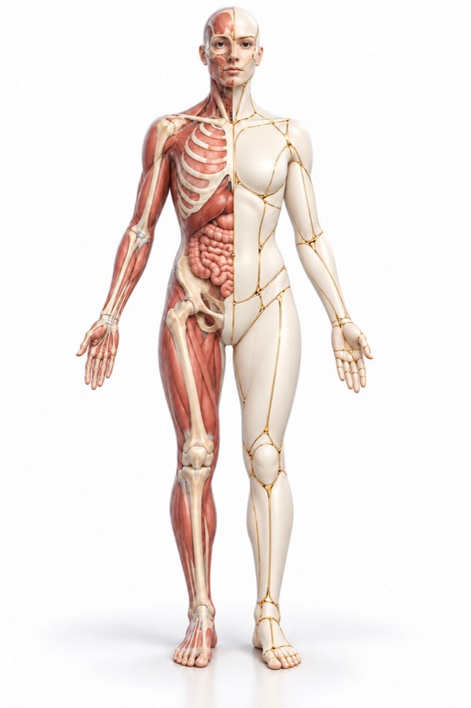
Conceptual visualization: restoration of a limb bone defect after injuryCONCEPTUAL VISUALIZATION
What it is
Part of the bone is missing after a wound, an open fracture, or removal of an infected bone.
How to restore
By the induced membrane method, fibula vascularized graft, or individual implant.
How long does it take
Several stages; in a meta-analysis of various methods, the bone healed on average in 7.8–8.9 months.
What comes first
Clean wound, bone covered with living tissue, and infection control.

What is
bone defect.

A bone defect is a gap in the bone that cannot be closed simply by putting fragments together. It can occur in the tibia, femur, humerus, and forearm, as well as the pelvis or skull. In studies of the induced membrane method (3840 patients), 60.9% of defects were in the tibia.

After modern injuries, defects can be large. In a series of 24 Ukrainian Armed Forces soldiers with humeral bone defects after gunshot wounds, 17 (71%) had a defect over 10 cm.

Bone restoration is not a single operation, but a plan: first, treat the wound and infection and cover the bone with living tissue, then restore the bone itself. After combat injuries, this is more complicated due to extensive soft tissue damage and early wound infection.

This is general information. The doctor will tell you what will suit you after the examination.

How
bone defect presents.

The bone defect is visible on an X-ray, but a person notices something else: the limb cannot bear weight, the wound is not healing, the leg or arm becomes shorter.

01

The limb cannot bear weight

You cannot put weight on the leg or lift a weight with the arm; the limb moves where it should not move.

02

Shortening or deformity

The leg or arm has become shorter or healed at an angle.

03

Wound or fistula

There is a wound over the bone that does not heal, or an opening from which pus is discharged. These are signs of infection after a fracture.

Fire wound
04

Signs of bone infection

Pain, swelling, redness, and warmth over the bone, high fever.

05

Lack of tissue

The bone or plate is visible in the wound or is covered only by thin skin. If the wound has opened after a recent surgery — see the surgeon who performed the operation immediately.

Soft tissue defect
06

Between stages

After hospitalization, there is a cement spacer or external fixation device in the bone, and the next surgery needs to be planned.

Where does it come from?
bone defect.

The injury itself destroys the bone, and infection can remove even more — in that case, the affected bone is removed.

Injury

High-energy weapons shatter the bone into many fragments and leave large defects — this is how war injuries in Ukraine are described.

Open fracture

When the skin and muscles are damaged along with the bone. In a meta-analysis of 5 studies (454 patients) with severe open tibial fractures, early wound closure with tissue reduced the odds of infection (odds ratio 0.34).

Infection after fracture

A frequent and serious complication of injuries. If the infection has damaged the bone, the affected part is removed, leaving a cavity that needs to be restored.

Smoking

According to a meta-analysis of 7,516 cases (fractures, osteotomies, arthrodeses), in smokers, the risk that the bone will heal slowly or fail to heal is 2.2 times higher.

When to seek medical advice?

An old bone defect is a planned situation and usually does not require emergency care. Immediate action is needed in the case of fresh trauma, signs of sepsis, or a thrombus.

103 immediately if:

  • bleeding from the wound does not stop, despite pressing;
  • after an injury, bone protrudes from the wound;
  • after an injury, the leg is twisted or turned out;
  • the skin is cold and clammy from sweat;
  • pulse is rapid or weak;
  • breathing is difficult or rapid and shallow;
  • skin, lips, or tongue are blue, gray, pale, or mottled;
  • confusion or slurred speech;
  • sudden severe pain in any part of the body;
  • sudden shortness of breath;
  • pain in the chest or upper back;
  • very rapid heartbeat;
  • the person fainted.

Do not delay a visit to the doctor:

Immediately - to the surgeon who operated, and if it does not work - to the waiting room: after surgery, pain or swelling in a limb gradually increases; the wound has opened, revealing a spacer, plate, or bone. On the same day: pain, swelling, redness, and warmth over the bone; high fever or chills and general malaise; pus flowing from a wound or fistula; throbbing pain and swelling of one leg; coughing up blood. Planned: After hospitalization, if there is a defect or spacer, the bone is not healing, the limb is shorter or deformed — see a surgeon with CT scans and medical records to plan the recovery.

How to evaluate
bone defect.

  1. Conversation and documents

    When and how the injury occurred, which surgeries have already been performed, whether there was an infection; medical records, surgical notes, and hospital images.

  2. Review

    The surgeon assesses the skin and scars over the bone, fistulas, joint movements, sensation, and blood circulation of the limb.

  3. X-ray and CT scan

    They show the length of the defect, positions of fragments and fixators. Based on CT data, the surgery is planned, and a custom implant is made.

  4. Infection search

    Blood tests, and during surgery — cultures from deep tissues. Infection is confirmed, in particular, by a fistula, pus, or identical microbes in at least two tissue samples.

  5. Vessels and tissues

    Before bone transplantation, blood circulation in the leg, the area from which the bone is taken, and the areas where it will be transplanted are assessed.

How to restore
bone.

Many methods can restore a large bone defect, but none are optimal for everyone. The choice depends on the defect length, the bone, the condition of the tissues, and infection.

01

First, the wound and infection

Without this, the bone is not restored.

Cleansing
Dead tissues and infected bone are removed; sometimes several operations are needed for this.
Tissue coverage
The bone is covered with living tissue — a local or microsurgical flap.
What to expect
In an analysis of 50 studies (1530 patients) with bone defects in post-fracture infection, the infection was cured in 83% after the first planned treatment and in 94% by the end of follow-up; reinfection occurred in 8%, amputation in 3%. Most of these studies are retrospective.
02

Induced membrane method

Two stages with a break of several weeks.

First stage
A cement spacer, often with antibiotics, is placed in the defect. A membrane rich in blood vessels forms around it.
Second stage
The spacer is removed, and the membrane is filled with the patient's own bone chips. The authors of the analysis of 78 studies recommend performing the second stage after 6–8 weeks: a break of more than 8 weeks was associated with higher chances that the bone would not heal.
The results
In the same analysis (3840 patients, average defect 6.4 cm) the bone healed in 87.6%. In a review of 156 patients with shoulder and forearm defects, the bone healed on average in 5.5 months, complications occurred in 21%.
03

Fibula on vessels

How is it
A portion of the fibula along with the vessels is transplanted into the defect and the vessels are stitched under a microscope.
The results
According to a meta-analysis of 110 articles (2226 patients), the bone healed in 80.1%; complications occurred in 39.4% (fractures, non-unions, infections, thrombosis), repeat surgeries in 24.6%. In another meta-analysis (74 articles, 1781 patients) among defects after trauma, healing with such bone occurred faster — on average in 6.9 months.
04

Individual implants

Pelvis
Based on CT, a titanium implant is printed in the shape of the lost fragment. Observation results are promising, but the method is still new.
Skull
In a meta-analysis of 9 studies (942 patients), 3D-printed titanium implants had fewer complications than standard ones, but fluid accumulation under the implant occurred more often; the quality of the studies was low.
05

Other methods

Bone lengthening with a device
The bone is gradually "stretched" using an external fixation device. In cases of infected non-union of the tibia with a defect, the bone healed in 94.6% after this method and in 88.0% after membrane technique — the difference is not significant, data are limited.
Bone chips without a membrane
The patient’s own bone or donor bone. In a meta-analysis of 74 studies, among post-traumatic defects, spongy bone resulted in fewer complications and reoperations, although overall after bone grafts the bone initially healed in 75% of cases. What is suitable depends, among other things, on the cause of the defect.

How
we treat it.

Bone defects after trauma at the Valikhnovskyi Surgery Institute are treated under the direction of "Surgery of Gunshot Injuries." The clinic closes wounds and restores bone and the tissues above it; a staged plan is developed after examination and CT scan.

CLINIC SERVICE Surgery of gunshot injuries Bullet, shrapnel, and landmine injuries — from a fresh wound to restoration of tissue, form, and function. View service

What we do for this condition

  • Surgical treatment and closure of wounds — if the wound does not heal or is infected, the cause is treated first, and then the wound is closed.
  • Microsurgical flaps — an exposed bone is covered with a tissue complex from another part of the body, sewing the vessels under a microscope.
  • Induced membrane method — large defects of limb bones are restored in two stages with an interval of several weeks.
  • Bone flap with vessels — a fragment of the fibula is transplanted along with the vessels.
  • Individual 3D-printed titanium implants — for pelvic and cranial bones — shaped according to the lost fragment based on CT data.

Rostislav Valikhnovskyi

Plastic surgeon, candidate of medical sciences, founder of Dr. Valikhnovskyi Institute of Surgery.

How treatment works with us

  1. Photo onlineSend CT, X-ray, medical records, and wound photos — the doctor will preliminarily assess the defect.
  2. ConsultationExamination of the limb, tissues over the bone, and signs of infection; if necessary — a new CT.
  3. Plan of stagesWhich to do first — wound, infection, or bone — and by what method to restore the defect.
  4. OperationsWound treatment and closure, then bone restoration — with a membrane, vascularized bone, or implant.
  5. RecoveryMonitoring bone healing on images, rehabilitation, and gradual loading.

Common
questions

Short answers. What is right for you, the doctor will tell you during the consultation.

Can the bone grow back on its own?

Depends on the size of the defect and the condition of the tissues. Large defects after injuries are surgically reconstructed — with a membrane, vascularized bone, or an implant. What you need will be determined by the surgeon after CT.

How long does the treatment take?

There are several stages: wound, infection, then bone. In a meta-analysis of different methods, the bone healed on average in 7.8–8.9 months. The surgeon explains the exact plan in advance.

I have a cement spacer in my bone. Can I wait for the second stage?

It is not worth delaying for too long: in an analysis of 78 studies, a gap between stages of 8 weeks was associated with higher chances that the bone would not heal. See a surgeon to plan the second stage.

Will my leg hurt from which the fibula is taken?

Complications from the donor leg do occur: in a meta-analysis, they made up 10.7% of all complications. The surgeon will explain what to expect specifically for you.

Can I smoke during treatment?

It’s better to quit: according to a meta-analysis, in smokers the risk that the bone will heal slowly or not at all is 2.2 times higher, and healing takes longer.

Is it possible to restore a bone if it has an infection?

Yes, but first the infection is treated: the affected bone is removed, a spacer is placed, often with an antibiotic, and only then is the defect filled.

Do you treat military personnel?

Yes. Surgeries for servicemen of the Armed Forces of Ukraine are carried out by the clinic with funding from charitable foundations; the administrator will explain the details.

Sources

19 sources · meta-analyses, systematic reviews, consensus, and NHS
showCollapse
  1. Induced membrane technique for large bone defects: A systematic review and individual participant data meta-analysisMedicine (Baltimore), 2022doi:10.1097/MD.0000000000029292 ↗
  2. Masquelet’s induced membrane technique in the upper limb: a systematic review of the current outcomesJournal of Orthopaedics and Traumatology, 2025doi:10.1186/s10195-024-00815-w ↗
  3. Vascularized fibular grafts for the treatment of long bone defects: pros and cons. A systematic review and meta-analysisArchives of Orthopaedic and Trauma Surgery, 2023doi:10.1007/s00402-021-03962-5 ↗
  4. Union, complication, reintervention and failure rates of surgical techniques for large diaphyseal defects: a systematic review and meta-analysisScientific Reports, 2022doi:10.1038/s41598-022-12140-5 ↗
  5. Distraction osteogenesis versus induced membrane technique for infected tibial non-unions with segmental bone loss: a systematic review of the literature and meta-analysis of available studiesEuropean Journal of Trauma and Emergency Surgery, 2024doi:10.1007/s00068-023-02375-w ↗
  6. Management of critical-sized bone defects in the treatment of fracture-related infection: a systematic review and pooled analysisArchives of Orthopaedic and Trauma Surgery, 2021doi:10.1007/s00402-020-03525-0 ↗
  7. Fracture-related infection: A consensus on definition from an international expert groupInjury, 2018doi:10.1016/j.injury.2017.08.040 ↗
  8. Management of severe defects of humerus in combat patients injured in Russo-Ukrainian warInjury, 2024doi:10.1016/j.injury.2023.111280 ↗
  9. Masquelet technique in military practice: specificities and future directions for combat-related bone defect reconstructionMilitary Medical Research, 2022doi:10.1186/s40779-022-00411-1 ↗
  10. Early orthoplastic coverage reduces fracture-related infections in Gustilo III B/C tibial fractures - a systematic review and meta-analysisEuropean Journal of Trauma and Emergency Surgery, 2026doi:10.1007/s00068-026-03189-2 ↗
  11. A systematic review of follow-up results of additively manufactured customized implants for the pelvic areaExpert Review of Medical Devices, 2023doi:10.1080/17434440.2023.2183839 ↗
  12. Meta-analyses of the surgical outcomes using personalized 3D-printed titanium and PEEK vs. standard implants in cranial reconstruction in patients undergoing craniectomyNeurosurgical Review, 2025doi:10.1007/s10143-025-03470-9 ↗
  13. Do smokers have greater risk of delayed and non-union after fracture, osteotomy and arthrodesis? A systematic review with meta-analysisBMJ Open, 2016doi:10.1136/bmjopen-2015-010303 ↗
  14. Broken legNHS, 2025nhs.uk/conditions/broken-leg ↗
  15. OsteomyelitisNHS, 2023nhs.uk/conditions/osteomyelitis ↗
  16. SepsisNHS, 2026nhs.uk/conditions/sepsis ↗
  17. Compartment syndromeNHS, 2026nhs.uk/conditions/compartment-syndrome ↗
  18. DVT (deep vein thrombosis)NHS, 2026nhs.uk/conditions/deep-vein-thrombosis-dvt ↗
  19. Pulmonary embolismNHS, 2026nhs.uk/conditions/pulmonary-embolism ↗

The figures on the page are from these sources.

VALIKHNOVSKI SURGERY PARTNERS

A defect remained
bones after injury?

Send the CT, reports, and photos. The doctor will say where to start and which method can restore the bone.

Call Send an enquiry
ONLINE ENQUIRY

Tell us how we can help.

Leave your contact details and briefly describe the situation. A coordinator will contact you to agree on the next step.

Preferred contact method