CONDITION / WOUNDS AND INJURIES

Soft tissue defect.

A soft tissue defect is an area where skin, fat, or muscles are missing after injury, trauma, infection, or surgery, and the wound does not close on its own. It is closed with a skin graft, dermal matrix, or flap — depending on what lies at the bottom of the wound.

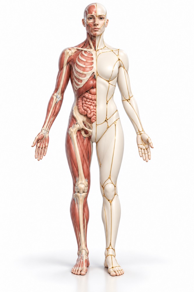
Conceptual visualization: closure of a soft tissue defectCONCEPTUAL VISUALIZATION
What it is
An area where skin, fat, or muscles are missing after injury, infection, or surgery.
What the method depends on
On what is at the bottom of the wound: muscles and fat, or exposed bone, tendons, vessels.
What to close
With skin, a dermal matrix with skin on top, or a flap — using one’s own tissues with blood supply.
Who treats it
A plastic surgeon, and if needed — a traumatologist, vascular surgeon, and rehabilitation specialist.

When tissue
is insufficient.

A common wound heals because the edges come together. When too much tissue is lost, the edges cannot be brought together without tension, and the wound remains open for weeks. Such an area is called a soft tissue defect.

According to the field of reconstructive leg surgery, such defects usually remain after injuries, infections, or tumor removal. Today the goal is not only to close the wound: function, appearance, and how the person evaluates the result also matter.

During the war, many such defects are caused by gunshot and blast injuries: large wounds with tissue loss and contamination, sometimes over bones or vessels. Patients seek help both with an open wound and after healing — when there is still a depression or a scar that tightens the skin.

This is general information. The doctor will tell you what will suit you after the examination.

How does it look
defect.

A defect is not a diagnosis of a single cause, but a condition of a wound or area. What is visible at the base determines how it will be closed.

01

The wound does not close

After cleaning, there remains an open area without skin, which does not decrease for weeks.

Fire wound

02

Exposed structures

At the bottom of the wound, tendons, bone, vessels, or metal structures are visible — they cannot be covered with skin alone.

03

Retraction

The wound has healed, but a depression remains at the site of lost fat and muscle, and the contour has changed.

05

The scar cracks

A thin scar over a bone is easily injured and cracks; an ulcer on it that does not heal is a reason to see a surgeon in the coming weeks.

Unstable scar

Where does it come from?
defect.

The cause affects how contaminated the wound is and how many stages of cleaning will be needed before closure.

Injury

In conflicts in Iraq and Afghanistan, limb injuries accounted for over 60% of all wounds, mostly from explosive devices. Such wounds combine extensive tissue loss and contamination.

Fire wound

Burns

A deep burn destroys all layers of the skin; such wounds are closed with skin grafts or dermal substitutes.

Post-burn contracture

After surgery

A section of skin has died or the wound has reopened after surgery, including after tumor removal.

Skin necrosis after surgery

When to seek medical advice?

An old defect or retraction after healing is a planned situation; they usually do not require urgent help. But an open wound and a postoperative wound can become complicated by bleeding or infection.

103 immediately if:

  • bleeding from the wound does not stop, despite pressing;
  • the blood is gushing, bright red;
  • breathing is very frequent;
  • confusion or slurred speech;
  • Skin or lips are blue, pale, or blotchy.

Do not delay a visit to the doctor:

Immediately - to the surgeon who operated, and if it does not work - to the waiting room: After flap transplantation, it has changed color or become cold, or pain and swelling in the operation area are increasing. On the same day: The wound is swollen, red, increasingly painful, or pus is coming out; you generally feel unwell or have a fever. In the coming weeks: The wound does not heal or is oozing; tendons or bone are visible; an ulcer that does not heal has appeared on the scar. Planned: Retraction, a scar that tightens the skin or is easily injured — consult a surgeon.

How to evaluate
defect.

  1. Conversation and documents

    What caused the defect, which operations have already been done, whether there are chronic diseases; discharge summaries, operation reports, and imaging.

  2. Examination of the wound

    Size and depth, what is at the base, whether there are signs of infection, the quality of the surrounding skin, circulation, and movement.

  3. Pictures

    X-ray or CT — if there is a defect in the bone, fragments, or metal structures.

  4. Wound culture

    If the wound is infected, material is taken from it to identify bacteria and select an antibiotic.

  5. Donor plots

    The surgeon assesses where skin or a flap can be taken and how this will affect that area.

How to close
defect.

The method is traditionally selected according to the principle of the reconstructive ladder — from simple to complex; a modern review emphasizes that it's important not only to close the wound, but also its function and appearance. The review of hand injuries first describes stabilization, then staged cleaning, and then closure according to the size and depth of the defect.

01

Wound preparation

Until the wound is clean.

Cleansing
Dead tissue and dirt are removed, if necessary, several times. An infected wound is usually not closed immediately.
Vacuum dressing
Between stages of wound cleaning, they can be kept under vacuum. In a study of 69 soldiers with limb vascular injuries and large defects, after vacuum treatment, wounds were sutured in 75.4%, covered with a skin graft in 17.4%, and with a flap in 4.3%.
02

Skin grafting

When at the bottom — muscles or fat.

How do they do it?
A thin layer of the patient’s own skin is taken from another area and placed on the clean wound. A split-thickness graft takes part of the skin’s thickness and covers large areas, while a full-thickness graft uses its entire thickness.
Vacuum on top
According to a meta-analysis of 16 randomized studies, a vacuum dressing on a grafted skin resulted in 8.3% higher graft survival than a standard dressing and fewer reoperations.
03

Dermal matrix

Artificial base under the skin.

How it works
A matrix is placed on the wound, blood vessels grow into it, and new dermis forms; then thin skin is grafted on top. These are two stages.
What is known
In a systematic review of 24 studies (202 patients) of synthetic matrix, almost complete (over 95%) matrix integration was observed in 84% of patients; the median time to skin grafting was 34 days. The authors note that the samples are small.
scar
According to a meta-analysis in burns, with one type of matrix the wound initially epithelialized more slowly than with skin alone, while with another type the subjective assessment of scar quality was better after 6 months.
04

Patches

When bone, tendons, vessels are exposed.

Local and perforator
Tissues adjacent to the defect are moved on a single vessel. According to a meta-analysis on defects of the lower third of the leg, flap loss occurred in 2.77% of propeller flaps and 3.9% of free flaps — the difference is not significant; partial necrosis occurred more frequently in propeller flaps: 6.88% versus 2.70%.
Experience in Ukraine
In a Ukrainian study, 62 wounded individuals with shrapnel defects of the legs (non-randomized) received pedicled flaps along with vacuum therapy, which was associated with a lower rate of infectious complications — 18.75% versus 30% — and a hospital stay shorter by 7 days.
Free flaps
Tissue complexes from the thigh or back are transplanted by suturing vessels under a microscope. In a systematic review of combat limb injuries (501 flaps), the average survival rate of the flaps was 95.5%.
Deadlines
According to a meta-analysis of 43 studies on leg trauma, a free flap performed within the first 72 hours resulted in fewer flap losses and infections than a delayed flap; in practice, most flaps are done later — after wound cleansing and treatment of more serious injuries.
05

After surgery

Follow-up
According to a systematic review of 44,031 free flaps, a repeat operation due to circulatory disorders was required in 5.8%. Among those reoperated on the first day, 93.8% of flaps were salvaged, by the third day — only 12.1%; therefore, the first 48 hours are monitored particularly closely.
Contour
Depressed areas after healing can be leveled with autologous fat grafting; the effect is assessed individually.

How
we treat it.

Soft tissue defects at the Doctor Valikhnovskyi Institute of Surgery are treated in the "Gunshot Wound Surgery" department; large defects after burns and multiple surgeries — together with the "Burn Reconstruction" and "New Body" departments.

CLINIC SERVICE Surgery of gunshot injuries Bullet, shrapnel, and landmine injuries — from a fresh wound to restoration of tissue, form, and function. View service

What we do for this condition

  • Surgical treatment and vacuum dressings (NPWT) — the wound is cleaned again and prepared for closure.
  • Skin grafting — split-thickness or full-thickness graft — for a wound without exposed tendons and bones.
  • Dermal matrices — two-layer or single-layer artificial dermis, and after vascular ingrowth — a thin layer of own skin.
  • Local flaps and Z-plasty — for small defects and scars that contract the skin.
  • Perforating flaps — tissue on a single vessel, in particular ALT thigh flap, — preserving the muscles of the donor site.
  • Free microsurgical flaps — ALT flap, latissimus dorsi flap; several flaps are sometimes transplanted in one operation.
  • Lipofilling — transplantation of own fat into sunken areas and scars.

Rostislav Valikhnovskyi

Plastic surgeon, candidate of medical sciences, founder of Dr. Valikhnovskyi Institute of Surgery.

How treatment works with us

  1. Photo onlineSend a photo of the defect, medical reports, and images — the doctor will preliminarily assess the situation.
  2. ConsultationExamination of the wound, assessment of blood circulation, movements, and donor areas.
  3. PlanWhether further cleaning is needed, how to cover the defect, and approximately how many stages.
  4. SurgerySkin transplantation, matrix or flap; after large reconstructions — several days in the hospital with monitoring of blood flow in the flap.
  5. RecoveryDressings, care of the donor site, movement rehabilitation; scars are corrected after maturation.

Common
questions

Short answers. What is right for you, the doctor will tell you during the consultation.

Why can't you just stitch up a wound?

When there is a lack of tissue, the edges cannot be brought together without tension: the suture will come apart or impair blood circulation. In that case, the wound is closed with skin, a matrix, or a flap.

Where is the skin for transplantation taken from?

Usually from areas covered by clothing — for example, from the thigh. A thin layer is removed so that the donor site heals on its own; a full-thickness graft is taken from areas that can be sutured.

Which is better: skin graft or flap?

It depends on what is at the base of the wound. Skin takes to muscles and fat; if bone, tendons, or vessels are exposed, a flap or a matrix is needed.

Why use a dermal matrix if you can transplant skin immediately?

The matrix creates a thicker layer under the skin where there is not enough skin itself. This is an additional step and time, so the surgeon uses it as indicated.

How many operations will be needed?

From one for a small defect to several for a contaminated or large wound: first cleaning, then closure, later — scar and contour correction.

Will the flap take?

In a systematic review of combat limb injuries, on average, 95.5% of free flaps survived, meaning there is a risk of loss. Therefore, the flap is carefully monitored during the first days.

The wound has already healed, but a depression remains. Can it be corrected?

Yes, this is a planned surgery: the depression is leveled with your own fat, a flap, or scar excision. The surgeon will say what is suitable after the examination.

Sources

14 sources · meta-analyses, systematic reviews, experience of the war in Ukraine and NHS
showCollapse
  1. Revisiting the reconstructive ladder for soft tissue reconstruction in the lower extremityAnnals of Translational Medicine, 2024doi:10.21037/atm-23-1445 ↗
  2. Changing paradigms in lower extremity reconstruction in war-related injuriesMilitary Medical Research, 2016doi:10.1186/s40779-016-0080-7 ↗
  3. Soft Tissue Reconstruction of the Upper Extremity After Ballistic InjuryHand Clinics, 2025doi:10.1016/j.hcl.2025.03.005 ↗
  4. Negative pressure wound therapy for combat-related extremity vascular injuries: clinical experience from the war in UkraineScandinavian Journal of Trauma, Resuscitation and Emergency Medicine, 2025doi:10.1186/s13049-025-01502-3 ↗
  5. Treatment of combat gunshot shrapnel trauma of large defects of the soft tissues of the lower extremities with plastic surgery in combination with vacuum assisted wound closure (VAC)Wiadomości Lekarskie, 2023doi:10.36740/WLek202305205 ↗
  6. Benefits of negative pressure wound therapy in skin grafts: A systematic review and meta-analysis of randomised controlled trialsJournal of Plastic, Reconstructive & Aesthetic Surgery, 2025doi:10.1016/j.bjps.2025.01.036 ↗
  7. Systematic Review and Meta-analysis of Biodegradable Temporizing Matrix Application for Complex Wound ReconstructionJournal of Burn Care & Research, 2025doi:10.1093/jbcr/irae081 ↗
  8. Outcomes of dermal substitutes in burns and burn scar reconstruction: A systematic review and meta-analysisWound Repair and Regeneration, 2024doi:10.1111/wrr.13226 ↗
  9. Free versus perforator-pedicled propeller flaps in lower extremity reconstruction: What is the safest coverage? A meta-analysisMicrosurgery, 2018doi:10.1002/micr.30047 ↗
  10. Free-Tissue Transfer for the Reconstruction of War-Related Extremity Injuries: A Systematic Review of Current PracticeMilitary Medicine, 2016doi:10.7205/MILMED-D-15-00059 ↗
  11. Meta-analysis of Timing for Microsurgical Free-Flap Reconstruction for Lower Limb Injury: Evaluation of the Godina PrinciplesJournal of Reconstructive Microsurgery, 2018doi:10.1055/s-0037-1621724 ↗
  12. Free Flap Monitoring, Salvage, and Failure Timing: A Systematic ReviewJournal of Reconstructive Microsurgery, 2021doi:10.1055/s-0040-1722182 ↗
  13. Cuts and grazesNHS, 2026nhs.uk/conditions/cuts-and-grazes ↗
  14. SepsisNHS, 2026nhs.uk/conditions/sepsis ↗

The figures on the page are from these sources.

VALIKHNOVSKI SURGERY PARTNERS

The wound does not close
Is there still a depression?

Send a photo of the defect, medical reports, and images. The doctor will say how it can be covered and how many stages will be needed.

Call Send an enquiry
ONLINE ENQUIRY

Tell us how we can help.

Leave your contact details and briefly describe the situation. A coordinator will contact you to agree on the next step.

Preferred contact method