Wound or lack of skin
After the tumor is removed, there is an area that needs to be closed, or it has healed with an indented scar.
 Discuss your case
Facial defects after oncological surgery — loss of skin, soft tissue, cartilage, or bone that remains after tumor removal. They are restored with their own tissues or epithesis — immediately or later, depending on what histology shows.
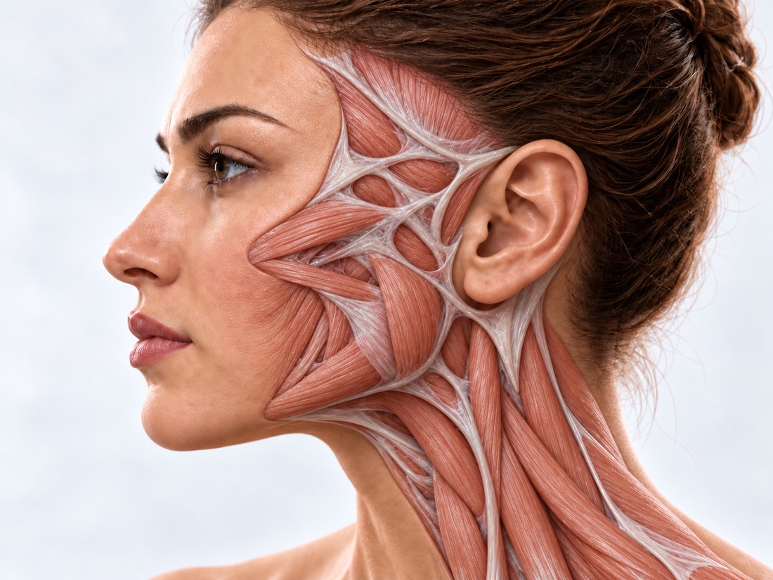 CONCEPTUAL VISUALIZATION
The main thing in the treatment of skin cancer is to remove the tumor completely. For squamous cell carcinoma of the skin, the European guideline calls clean incision edges the most important condition for surgery. Therefore, together with the tumor, the surgeon takes away a supply of healthy tissues, and a defect remains on the face.
The defect may be small—a few centimeters of skin on the cheek or nose—or involve cartilage, bone, eyelid, lip, or auricle. Then it is not only the appearance that suffers: the eye may close more poorly, the lip - more difficult to close, the nose - more difficult to breathe.
The goal of reconstruction is to bring the face closer to what it was before the disease and preserve function. If cancer treatment is still ongoing, reconstruction is planned together with the oncologist.
Changes in the face after surgery significantly affect the quality of life. The support of loved ones and specialists and clear information about treatment help to adapt to this.
This is general information. The doctor will tell you what will suit you after the examination.
The consequences depend on where the tumor was and how much tissue had to be removed.
After the tumor is removed, there is an area that needs to be closed, or it has healed with an indented scar.
Missing part of wing, tip or back; the nose breathes worse.
Nose defects after removal of tumorsThe scar pulls the eyelid down, it turns out or the eye does not close completely.
EctropionThe lip is deformed, the mouth opens or closes worse.
Lip defectsPart of the auricle is missing after tumor removal.
Loss of auricleAfter major surgery or radiation therapy, the cheek sinks, the tissues are compacted, the face is asymmetrical.
How and when the face is restored determines not only the size of the defect.
Basal cell carcinoma is the most common malignant tumor in fair-skinned people. High-risk tumors and those that have returned are advised to be removed with microscopic margin control.
Basal cell carcinoma of the faceUntil histology has confirmed that the tumor has been completely removed, complex reconstruction is often postponed.
Irradiated tissue heals worse: in a review of 53 studies, complete loss of the free flap in the irradiated area occurred about 1.6 times more often.
Whether the cartilage, bone, eyelid, lip, or nerve is affected — this depends on the choice of flap and the number of stages.
A long-standing defect after an operation that has already healed is a planned situation: it usually does not require emergency care. In the first weeks after surgery, some symptoms require immediate help.
Immediately - to the surgeon who operated: in the first two weeks after the operation, the flap changed color (darkened, blue or pale), became cold to the touch or tight and tense; if it is not possible to contact the surgeon, go to the reception department. On the same day: the wound is swollen, reddened, it hurts more and more, pus flows from it, you feel bad or have a high temperature. Planned: scars, asymmetry, deformity of the nose, eyelids or lips after healing. If a new nodule, ulcer, or thickening appears at the site of the operation or nearby, tell the oncologist without waiting for the next control examination.
An extract about the operation, histology with a conclusion about the edges of the incision, data about radiation therapy; photo of the face before the disease, if any.
The size and depth of the defect, the condition of the skin nearby, scars; how the eye closes, the lips close, the nose breathes.
If bones or deep tissues are affected; bone reconstruction is planned according to the CT scan.
Whether the treatment of the tumor is completed, whether radiation therapy is needed and how often to come for control.
The method is chosen according to the size, location and depth of the defect, the condition of the tissues after treatment and the wishes of the patient. Some defects are closed in one operation, complex ones - in several stages.
The main way.
When tissue reconstruction is impossible or undesirable.
Facial defects after oncological operations are restored at Dr. Valikhnovskyi's Institute of Surgery in the "New Face" direction. Reconstruction is often done in one operation with removal of the tumor, if the edges of the incision are clean; otherwise - after histology. The clinic is responsible for removal and reconstruction; Chemo-, immuno-, and radiation therapy and oncological supervision are conducted by an oncologist.
Plastic surgeon, candidate of medical sciences, founder of Dr. Valikhnovskyi Institute of Surgery.
Short answers. What is right for you, the doctor will tell you during the consultation.
Often yes, if histology confirms that the tumor has been completely removed. If the edges of the incision have not yet been checked, the complex reconstruction is postponed.
Therefore, they are not in a hurry with a complex reconstruction until clean edges have been confirmed. After the reconstruction, regular check-ups with the oncologist continue; show new nodules or ulcers to the doctor without waiting for a routine examination.
Yes, but irradiated tissues heal worse, and the risk of complications is higher. This is taken into account when choosing a method.
Depends on the defect, tissue condition and general health. Own tissues become part of the face; epithesis helps when reconstruction is impossible or undesirable. Decisions are made together with the patient.
A small defect is often closed in one operation. Large ones - in several stages, and scars and contour may need correction later.
It is possible: the scars are cut out, the contour is adjusted with fat, the deformity is corrected by repeated surgery. What is possible for you, the doctor will tell after the examination.
The figures on the page are from these sources.
Send a photo of the face, an extract and a histology. The doctor will tell how and when the tissue can be restored.
Leave your contact details and briefly describe the situation. A coordinator will contact you to agree on the next step.