CONDITION / FACE

Dermoid cyst of the face and lipodermoid.

A dermoid cyst is a congenital benign cyst, most often near the outer edge of the eyebrow, that is usually noticed in childhood. It is removed completely, and cysts in the middle of the nose or forehead are examined by MRI or CT before surgery: they can reach the skull.

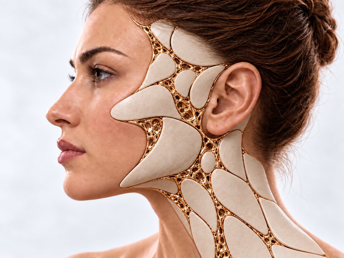
Conceptual visualization: eyebrow, eye and nose areaCONCEPTUAL VISUALIZATION
What it is
Congenital benign cyst, the wall of which is similar to the skin.
Where it happens
Most often near the eye: in a series of 49 children, 61% of head and neck dermoid cysts were around the orbit.
When noticed
Usually in early childhood; part - immediately after birth.
Which helps
Complete removal; median nose and forehead cysts — after MRI or CT scan.

What is
dermoid cyst.

A dermoid cyst is a congenital benign formation. It is laid even before birth where the tissues of the face grow: a piece of skin gets deep and slowly forms a cyst. Therefore, the wall of the cyst is similar to the skin, and the horny substance accumulates inside.

Most often, a dermoid cyst is near the outer corner of the eye, under the edge of the eyebrow - doctors call it external corner. Less often - near the inner corner of the eye, on the bridge of the nose, in the middle of the nose, on the forehead, temple or scalp. Dermoid cysts occur more often in children, but they also occur in adults.

Lipodermoid, or dermolipoma, is a related congenital formation on the surface of the eye, under the eyelid: a soft tissue made of fat, covered with a dermis. About a third of lipodermoids have hairs.

These formations themselves are benign. But the cyst can grow slowly, push the bone and become inflamed, and cysts in the middle of the nose and forehead sometimes have a course inside the skull. Therefore, they are examined and usually removed.

This is general information. What will suit you or your child, the doctor will tell after the examination.

How
dermoid cyst presents.

Most often, parents notice a smooth ball near the eyebrow in a child. Other signs depend on where the cyst lies.

01

A bump that doesn't hurt

A dense, smooth lump under the skin near the eyebrow or eye. In a series of 203 patients, 94% of cysts looked like this.

02

Movable or attached

The cyst can move under the skin or be tightly attached to the bone - the doctor evaluates this during the examination.

03

Nose hole

In the middle of the nose there is a tiny pit or hole, from which the contents are sometimes released. This is a move that can lead deep.

Congenital defects of the nose
04

Signs from the side of the eye

Less often, the eye protrudes slightly, shifts, squints or sees worse. Such changes occur more often in adults than in children.

05

Lipodermoid

Soft formation on the surface of the eye under the eyelid. Most often it is noticed because it increases over the years; can irritate the eye, cause redness and mucous discharge.

06

Inflammation

The cyst suddenly becomes red, swollen and painful - for example, if it has burst.

Why does it occur?
and what threatens

A dermoid cyst is a developmental defect, not the result of something done by the parents or the person himself. Another thing is more important: how it behaves over time.

Congenital defect

The cyst is formed where facial tissues grow. In a series of 117 patients with cysts of the nose, forehead and head, half of the cysts were noticed before 4.8 months, and in 42 cases - immediately after birth.

Pressure on the bone

The cyst grows slowly and can push into the bone: in a series of 280 cysts near the eye, bone changes were present in the majority of patients.

A cyst through the bone

Part of the cyst near the eye passes through the bone into the temporal fossa like a dumbbell: in the same series — 16 (5.7%).

Connection with the skull

Cysts in the middle of the forehead and scalp more often extend into the skull than on the sides: 54.5% versus 17.5%. There is a high risk of such a course — and in cysts of the nose and temporal area.

Combination with other defects

Lipodermoid is sometimes combined with hemifacial microsomia — underdevelopment of one half of the face: in a series of 34 patients, 10 had this combination.

Hemifacial microsomia

When to seek medical advice?

A dermoid cyst is a planned situation, it usually does not require emergency care. A rare danger is infection of a cyst in the middle of the nose or forehead that communicates with the skull cavity: it can cause meningitis or a brain abscess. An inflamed cyst near the eye can cause an infection of the eye socket. Call 103 if at least one of the following occurs:

103 immediately if:

  • very high or very low temperature, fever or severe chills;
  • very strong headache;
  • it is difficult to bend the neck, and it is painful to look at a bright light;
  • a rash that does not fade if a glass is pressed to it;
  • first seizures in life;
  • confusion or slurred speech;
  • the child is lethargic, difficult to wake up, does not eat or cries shrill or continuously;
  • after surgery near the eye or with an inflamed cyst near the eye, pain increases in or around the eye, the eye protrudes, it hurts when moving the eyes, or the eyelids do not open;
  • after an operation near the eye or with an inflamed cyst near the eye, double vision appeared or worsened.

To the doctor on the same day, if:

On the same day: the cyst is red, swollen, hot, or painful; pus flows from it or from the opening on the nose; the swelling has spread to the eyelid. In the coming days: the cyst grows noticeably, the eye gradually, without redness and swelling, began to protrude or itch, the lipodermoid irritates the eye or gives discharge. Planned: a bump near the eyebrow or on the nose that was noticed in a child - make an appointment for a consultation; it is better to remove the cyst without waiting for complications.

As defined,
or a deep cyst.

  1. Discussion and review

    When the bump was noticed, whether it was growing or inflamed. The doctor evaluates the location, size and mobility of the cyst, looks for the opening on the nose and signs from the side of the eye.

  2. A typical cyst near the eyebrow

    If the cyst is near the outer edge of the eyebrow and there are no other signs, imaging is usually not necessary: in a study of 61 children, 19 children had imaging, and none of the imaging showed progression into the eye socket or skull and did not alter treatment.

  3. MRI or CT

    Necessary for cysts in the middle of the nose, forehead and head, in the temporal area, with an opening with secretions or signs from the side of the eye. MRI more accurately shows the connection to the skull: in a study of 117 patients, it did not miss any such moves, while CT detected 72.7% of such moves.

  4. Ophthalmologist's examination

    If there is a lipodermoid, the cyst lies in the eye socket or there are changes in the eye.

How we treat
dermoid cyst.

The main treatment is to remove the cyst completely, together with the wall. The scope of the operation depends on where the cyst lies and whether it goes deep.

01

A cyst near the eyebrow and eye

The most common option.

Complete removal
The cyst is removed whole. After complete removal, relapses are rare: in a series of 49 children, the cyst returned in one.
Do not delay
The authors of the series of 280 cysts advise removing dermoid cysts early: most had already had bone changes, and 25% had signs of chronic inflammation in the cyst tissue.
Where is the incision made?
The incision can be hidden in the fold of the upper eyelid or a small incision can be made above the cyst. In a study of 47 children with a cyst near the inner corner of the eye, both approaches produced a better scar than an incision under the eyebrow; in the fold of the eyelid, the scar was the least noticeable.
If the cyst bursts
This happens during surgery, and long-term inflammation afterward is rare. After spontaneous rupture, the inflammation is prolonged more often.
02

A cyst in the middle of the nose and forehead

May have a course to the base of the skull.

Examination before surgery
An MRI or CT scan shows whether the cyst is deep and helps to plan the operation.
Removal of the cyst and course
The cyst and passage are removed completely. Access depends on whether it is a cyst or a process, where it lies and whether there is a continuation into the skull. If it is, a neurosurgeon takes part in the operation.
Restoring the shape of the nose
If the shape changes after removal, the back or tip of the nose is restored - with auricle cartilage, fascia or own fat.
Why not delay
An infected cyst that connects to the skull can cause meningitis or a brain abscess.
03

Lipodermoid

Surgery is not always necessary.

Follow-up
If the lipodermoid does not bother you, it can be observed: in a series of 120 patients, 40 (33%) decided to do so.
Surgery
It is removed when it irritates the eye, gives a discharge or noticeably increases: the changed cover is removed together with the fatty part and the wound is closed with conjunctiva.
Outcome and risks
After the operation, the complaints disappeared in all but one patient; rarely there are conjunctival adhesions or a small limitation of lateral movement of the eye.

How
we treat it.

Dermoid cysts of the face are removed in the "New face" direction at Dr. Valikhnovskyi's Institute of Surgery. The clinic is responsible for cyst removal and tissue restoration. If the cyst reaches the skull cavity, this part of the operation is conducted by a neurosurgeon; surgery on the surface of the eye for lipodermoid is planned together with an ophthalmologist.

CLINIC SERVICE A new face Form, symmetry, facial expressions and functions after injuries, wounds, burns, removal of tumors and congenital changes. View service

What we do for this condition

  • Removal of skin and soft tissue formations — the cyst is removed whole, together with the wall; removed is examined under a microscope.
  • Access through the eyelid crease — the incision is hidden in the natural crease of the upper eyelid, as during blepharoplasty.
  • Restoration of bone and contour — if the cyst has pushed through the bone of the forehead or the edge of the eyeball, the contour is restored with one's own bone or an implant.
  • Local patches — if a skin defect remains on the nose or forehead after removal of the cyst and procedure, it is covered with tissues from the adjacent area.

Rostislav Valikhnovskyi

Plastic surgeon, candidate of medical sciences, founder of Dr. Valikhnovskyi Institute of Surgery.

How treatment works with us

  1. Photo onlineSend a photo of the lump directly and from the side - the doctor will first assess the situation.
  2. ConsultationReview; if necessary, referral to an MRI or CT scan and to an ophthalmologist.
  3. PlanAccess and scope of the operation — taking into account where the cyst lies and whether it goes deep.
  4. SurgeryChildren are usually under general anesthesia; the cyst is removed completely.
  5. RecoveryThe doctor will explain how to care for the seam and when to come for an examination; result of histology.

Common
questions

Short answers. What is right for you, the doctor will tell you during the consultation.

The child has a lump near the eyebrow. Is it dangerous?

Most often, this is an external angular dermoid cyst. It is benign and rarely connects to the eye socket or skull. But the cyst grows and can push the bone, so it is better to show it to the surgeon.

Why remove if the cyst does not hurt?

Removal is the main treatment for dermoid cysts. In a series of 280 cysts near the eye, the majority already had bone changes, and 25% had signs of chronic inflammation in the cyst tissue; depending on the location, the cyst may extend into the skull.

Are MRIs or CT scans necessary before surgery?

For a typical cyst near the outer edge of the eyebrow, usually not. For cysts in the middle of the nose, forehead and head, in the temple, with an opening with secretions or changes from the side of the eye - yes.

At what age is the operation performed?

In a series of 117 patients with cysts of the nose, forehead, and head, half of the cysts were removed by 1.8 years. The doctor determines the terms individually - according to the size and location of the cyst.

Will the cyst return?

After complete removal, relapses are rare: in a series of 49 children, the cyst returned in one.

Will there be a scar?

A small scar remains. Near the eye, the incision can be hidden in the fold of the upper eyelid - so the scar is the least noticeable.

How does a lipodermoid differ from a dermoid cyst?

A dermoid cyst is a closed ball under the skin. Lipodermoid is a soft formation made of fat on the surface of the eye under the eyelid. Lipodermoid does not always need to be operated.

What will happen if the cyst bursts?

Long-term inflammation after a tear during surgery is rare. After spontaneous rupture, it is prolonged more often, so it is better to remove the cyst in advance.

Sources

17 sources · reviews, guidelines and research
showCollapse
  1. Orbital and periorbital dermoid/epidermoid cyst: a series of 280 cases and a brief reviewCanadian Journal of Ophthalmology, 2020doi:10.1016/j.jcjo.2019.08.005 ↗
  2. Orbital and periorbital dermoid cysts: Comparison of clinical features and management outcomes in children and adultsEuropean Journal of Ophthalmology, 2021doi:10.1177/1120672120964686 ↗
  3. Pediatric dermoid cysts of the head and neckOtolaryngology–Head and Neck Surgery, 2005doi:10.1016/j.otohns.2005.03.005 ↗
  4. Epidermoid and Dermoid Cysts of the Head and NeckJournal of Craniofacial Surgery, 2021doi:10.1097/SCS.0000000000006834 ↗
  5. Craniofacial Epidermoid and Dermoid CystsJournal of Craniofacial Surgery, 2023doi:10.1097/SCS.0000000000009561 ↗
  6. Diagnosing Common Benign Skin TumorsAmerican Family Physician, 2024pubmed.ncbi.nlm.nih.gov/39418568 ↗
  7. Is pre-operative imaging of external angular dermoid cysts in children necessary?Journal of Paediatrics and Child Health, 2022doi:10.1111/jpc.16008 ↗
  8. Imaging Yield and Surgical Outcomes of Nasal, Medial Brow, Forehead, and Scalp Dermoid CystsCleft Palate–Craniofacial Journal, 2026doi:10.1177/10556656241295572 ↗
  9. Clinical Outcomes of Ruptured Periorbital and Orbital Dermoid CystsOphthalmic Plastic and Reconstructive Surgery, 2017doi:10.1097/IOP.0000000000000735 ↗
  10. Comparison of three surgical techniques for internal angular dermoid cysts: a randomized controlled trialEye, 2022doi:10.1038/s41433-021-01851-0 ↗
  11. Nasal dermoidsCurrent Opinion in Otolaryngology & Head and Neck Surgery, 2006doi:10.1097/MOO.0b013e32801042db ↗
  12. Paediatric nasal dermoid: evaluation and managementCurrent Opinion in Otolaryngology & Head and Neck Surgery, 2021doi:10.1097/MOO.0000000000000765 ↗
  13. Intracranial complications of midline nasal dermoid cystsActa Chirurgica Belgica, 2019doi:10.1080/00015458.2017.1411552 ↗
  14. The Presentation and Surgical Treatment of Peribulbar DermolipomasOphthalmic Plastic and Reconstructive Surgery, 2021doi:10.1097/IOP.0000000000001848 ↗
  15. Prevalence of Oculo-auriculo-vertebral Spectrum in DermolipomaOphthalmology, 2013doi:10.1016/j.ophtha.2013.01.015 ↗
  16. MeningitisNHS, 2026nhs.uk/conditions/meningitis ↗
  17. CellulitisNHS, 2024nhs.uk/conditions/cellulitis ↗

The figures on the page are from these sources.

VALIKHNOVSKI SURGERY PARTNERS

Lump near the eyebrow
or on the child's nose?

Send a photo. The doctor will tell if examinations are needed and how the cyst can be removed.

Call Send an enquiry
ONLINE ENQUIRY

Tell us how we can help.

Leave your contact details and briefly describe the situation. A coordinator will contact you to agree on the next step.

Preferred contact method