CONDITION / EARS

Ear appendages and preauricular fistula.

Preauricular fistula is a tiny congenital hole in front of the ear, from which there is a narrow passage under the skin; an additional ear appendage is a congenital tubercle of the skin there. For the most part, they don't bother you, but the fistula can become inflamed, and then it is removed completely.

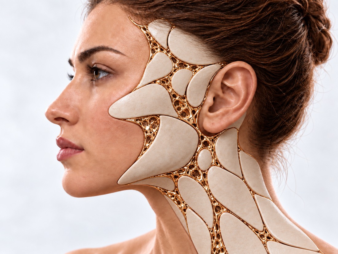
Conceptual visualization: auricle and area in front of the earCONCEPTUAL VISUALIZATION
What it is
A fistula is a hole in front of the ear with a narrow passage under the skin; an appendage is a lump of skin in front of the ear.
How common
In a survey of 23,533 people in Korea, fistula was unilateral in 1.3% and bilateral in 0.3%.
What bothers
Mostly nothing; fistula can fester and become inflamed.
How we treat
Without symptoms - observed; with repeated inflammations, the course is removed completely.

What is
fistula and appendix.

The auricle is formed before birth from six small tubercles. The most common explanation for a fistula is that these nodules have not fully fused. Then there is a tiny hole in the skin in front of the ear, from which a narrow passage, sometimes with a cyst, stretches under the skin. Doctors call it a preauricular fistula or sinus. The pit is usually near the front edge of the curl — the upper rim of the ear.

An additional auricular appendage (doctors say "accessory tragus") is a congenital tubercle of the skin in front of the ear, a defect in the development of part of the external ear. It occurs moderately rarely.

Both defects are benign. According to a review of the literature, a fistula is bilateral in 25–50% of cases, and in 3–10% it is one of the signs of another condition or syndrome, primarily deafness. Therefore, the doctor examines the child completely and, if necessary, checks the hearing and kidneys.

This is general information. What will suit your child, the doctor will tell after the examination.

How do they look?
fistula and appendix.

Usually, a dimple or bump does not bother the child. The reason to go to the doctor sooner is discharge or inflammation.

01

Pit in front of the ear

A tiny hole or depression near the upper front edge of the ear. Usually she doesn't bother with anything.

02

Allocation

Contents may be released from the hole. Among 121 young men with fistula, symptoms appeared in 24.0%, discharge was the most common, and they usually recurred.

03

Inflammation

The skin around the fistula reddens, swells and hurts, an abscess may form. Sometimes it appears not near the fossa itself, but lower on the cheek in front of the ear.

04

Lumpy skin

An additional appendage is a bump of skin in front of the ear, visible from birth.

05

Other features

Asymmetry of the face, defects of the eyelids or eyes, a small lower jaw, cysts or fistulas on the neck, an underdeveloped auricle - then the doctor will offer a wider examination.

Microtia

Why
they appear.

Both defects are congenital: they are formed during the intrauterine development of the ear.

Incomplete fusion

The six tubercles that make up the auricle have not fully fused - this is the most common explanation for the appearance of a fistula.

Heredity

Fistula can be accidental or inherited. In a study of 23,533 people, bilateral fistula in the father or mother was associated with bilateral fistula in the child; no such connection was found for one-sided.

Syndromes

Sometimes a fistula or appendix is part of the syndrome. For example, with a rare branchio-oto-renal syndrome, hearing and kidneys are also affected, and an additional goiter occurs, in particular, with Goldenhar syndrome.

Hemifacial microsomia

When to seek medical advice?

The fistula or appendix itself does not require emergency care. But a fistula can become infected, and an infection in a baby can quickly worsen the condition. Call 103 if a child or adult has at least one of the following:

103 immediately if:

  • swelling of the lips, tongue or throat, difficulty swallowing or speaking;
  • very frequent or difficult breathing - the baby grunts or pulls in the stomach during inhalation;
  • blue, gray, pale or marbled skin, lips or tongue (on dark skin it is more noticeable on the palms and feet);
  • skin is cold or sticky to the touch;
  • a rash that does not fade when pressed;
  • a child under 3 months has a temperature of 38 °C and above, at 3–6 months — 39 °C and above;
  • a child under 5 years of age has a temperature below 36 °C, is cold to the touch or shivers;
  • weak, shrill or continuous crying;
  • the child has fainted, it is difficult to wake him or he cannot be woken up;
  • a child under the age of 5 has not urinated for 12 hours;
  • convulsions;
  • a child over 5 years old or an adult has a very high or very low temperature or chills;
  • in a child from 5 years old or an adult - confusion, slurred speech or incomprehensible words.

Do not delay a visit to the doctor:

On the same day: the skin near the fistula or appendix is reddened, swollen, painful or hot to the touch; redness or swelling spreads; pus flows from the hole. Also show the doctor the abscess on the cheek in front of the ear - it may be associated with a fistula. Planned: a pit or bump near the ear is nothing to worry about, sometimes contents are released from the hole, the fistula has already become inflamed, or you do not know whether the child's hearing has been checked - make an appointment for a consultation.

How we assess
a child

  1. Review

    The doctor examines the fossa or appendage, both ears, and the face and looks for other abnormalities that may indicate the syndrome.

  2. hearing

    In a study of 68,484 newborns, permanent hearing loss occurred in 8 out of 1,000 babies with appendages or pits versus 1.5 out of 1,000 without them. Standard prenatal hearing screening has picked up all of these cases except for one late one, so it's important that it is done.

  3. Kidneys

    Ultrasound of the kidneys is recommended if there are other defects or features of the syndrome, deafness or ear or kidney defects in the family, diabetes in the mother during pregnancy. Without these signs, opinions differ — the doctor makes the decision.

  4. Before the operation

    The doctor finds out where and how far the stroke extends in order to remove it completely.

How we treat
fistula and appendix.

If the fistula does not bother you, and there are no other defects, according to the general opinion of experts, additional treatment is not necessary - it is enough to observe. Repeated inflammations are a clear reason to remove the fistula completely: this is the only way to get rid of it permanently.

01

Inflamed fistula

First, remove the infection.

Antibiotics
They are prescribed by a doctor; if necessary, a culture is taken from the secretions to select the drug.
Opening of the abscess
If an abscess has formed, it is opened and pus is released. But the course itself remains, so the inflammation can recur.
Do not miss the fistula
If the abscess appeared on the cheek in front of the ear, it is important to notice the tiny hole near the ear: otherwise the treatment will be incomplete and the operations will be unnecessary.
02

Surgery

Fistula: complete removal of the stroke.

How to delete
The course is removed together with the surrounding tissues, and not just the canal itself; augmentation during surgery and tracing the ramifications of the course may further reduce the risk of recurrence.
Return risk
In a review of 14 studies, the fistula returned in 5.5% after excision of the passage itself and in 2.2% after wider access above the ear; Excision of the course under the microscope gave 1.9%.
During inflammation
In a study of 212 patients, the results of surgery during and without active inflammation did not differ. The authors believe that it is possible to operate even when the inflammation does not respond well to treatment.
03

The appendix and associated conditions

Removal of the appendix
The appendix is usually removed surgically - this is the most common method, and the result is usually good.
Hearing and kidneys
If hearing loss or changes in the kidneys are found, they are treated by specialized doctors - an ENT doctor and a nephrologist.
Syndrome
In the case of branchio-oto-renal syndrome, the treatment is carried out together by an ENT doctor, a nephrologist and a geneticist.

How
we treat it.

Congenital ear defects are treated at Dr. Valikhnovskyi's Institute of Surgery in the field of "Surgery of congenital defects" along with the field of "Growing the Ear". The clinic is responsible for surgery and restoration of the shape of the ear; hearing is checked by an ENT doctor, kidneys by a nephrologist.

CLINIC SERVICE Surgery of birth defects Congenital defects of the face and body in children and adults — from the first operation to the correction of the consequences. View service

What we do for this condition

  • Microsurgical technique — magnifying optics and thin suture material help to accurately match the tissues and make the scar less noticeable.
  • Local flaps and cartilage — if a defect remains near the ear after removal, it is covered with tissues from the adjacent area.
  • Reconstruction of the auricle — if the defect in front of the ear is combined with underdevelopment of the ear — a frame made of costal cartilage or an implant.
  • Phased treatment as growth progresses — with syndromes such as hemifacial microsomia — jaw distraction, ear reconstruction, lipofilling.

Rostislav Valikhnovskyi

Plastic surgeon, candidate of medical sciences, founder of Dr. Valikhnovskyi Institute of Surgery.

How treatment works with us

  1. Photo onlineSend a photo of the child's ear from the side showing the fossa or appendage.
  2. ConsultationExamination of ears and face, conversation about inflammation, hearing and family history.
  3. ExaminationIf necessary, a hearing test by an ENT doctor and an ultrasound of the kidneys.
  4. The solutionWhether intervention is necessary and when - the doctor decides after the examination, taking into account inflammation, hearing and accompanying defects.
  5. ControlExaminations after treatment to detect the return or new inflammation in time.

Common
questions

Short answers. What is right for you, the doctor will tell you during the consultation.

Is it necessary to remove the fistula, if it does not bother?

Not necessarily. If the fistula has never been inflamed, and there are no other defects, it is enough to observe the common opinion of specialists. But among 121 young men with fistula, symptoms appeared in 24.0%, and in 7 out of 29 — already after 16 years.

Why does a fistula fester again after opening?

Opening only releases pus, and the course itself remains. You can finally get rid of a fistula only by complete excision.

Can a fistula return after surgery?

Maybe: in a review of 14 studies — in 5.5% after excision of the passage itself and in 2.2% after wider access above the ear.

Do I need a hearing test?

Yes, it is important for the baby to undergo a regular hearing screening. In a large study of newborns with appendages or pits, permanent hearing loss occurred more often — 8 out of 1,000 versus 1.5 out of 1,000. In other studies of children with isolated communication defects, no connection was found, so additional tests are ordered by the doctor as needed.

Is kidney ultrasound mandatory?

It is advised if there are other defects or features of the syndrome, deafness or ear or kidney defects in the family, diabetes in the mother during pregnancy. Without these signs, opinions differ, so the doctor makes the decision after the examination.

Is fistula transmitted to children?

Maybe: a fistula is inherited. Bilateral fistula in a child is associated with bilateral fistula in the father or mother.

When do you urgently need to see a doctor?

On the same day, if the skin near the ear is red, swollen, painful, hot, or pus flows from the hole. Call 103 if the child has difficulty breathing, a rash that does not fade, convulsions, a very high temperature (in a baby under 3 months — 38 °C and above) or other dangerous signs from the list above.

Sources

18 sources · reviews, research and NHS
showCollapse
  1. The preauricular sinus: a review of its clinical presentation, treatment, and associationsPediatric Dermatology, 2004doi:10.1111/j.0736-8046.2004.21301.x ↗
  2. The preauricular sinus: A review of its aetiology, clinical presentation and managementInternational Journal of Pediatric Otorhinolaryngology, 2005doi:10.1016/j.ijporl.2005.07.008 ↗
  3. Auricular fistula: a review of its clinical manifestations, genetics, and treatmentsJournal of Molecular Medicine, 2023doi:10.1007/s00109-023-02343-2 ↗
  4. A systematic review on the surgical outcome of preauricular sinus excision techniquesThe Laryngoscope, 2016doi:10.1002/lary.25829 ↗
  5. Preauricular sinus: clinical course and associationsArchives of Otolaryngology–Head & Neck Surgery, 2007doi:10.1001/archotol.133.1.65 ↗
  6. Analysis of incidence and genetic predisposition of preauricular sinusInternational Journal of Pediatric Otorhinolaryngology, 2014doi:10.1016/j.ijporl.2014.10.027 ↗
  7. Preauricular skin tags and ear pits are associated with permanent hearing impairment in newbornsPediatrics, 2008doi:10.1542/peds.2008-0606 ↗
  8. Isolated preauricular pits and tags: is it necessary to investigate renal abnormalities and hearing impairment?European Archives of Oto-Rhino-Laryngology, 2008doi:10.1007/s00405-008-0595-y ↗
  9. Is routine audiometric testing necessary for children with isolated preauricular lesions?International Journal of Pediatric Otorhinolaryngology, 2017doi:10.1016/j.ijporl.2016.12.032 ↗
  10. Syndromic ear anomalies and renal ultrasoundsPediatrics, 2001doi:10.1542/peds.108.2.e32 ↗
  11. Renal Ultrasound in Patients With Preauricular Skin Tags: Is It Necessary and Who Needs It?Plastic Surgery (Oakville), 2026doi:10.1177/22925503261419803 ↗
  12. Review of accessory tragus with highlights of its associated syndromesInternational Journal of Dermatology, 2014doi:10.1111/ijd.12369 ↗
  13. Surgical results of infected preauricular sinus: No need for delayInternational Journal of Pediatric Otorhinolaryngology, 2020doi:10.1016/j.ijporl.2020.110129 ↗
  14. Management of the infected preauricular sinusJournal of Plastic, Reconstructive & Aesthetic Surgery, 2023doi:10.1016/j.bjps.2023.05.046 ↗
  15. Branchio-oto-renal syndrome: a narrative reviewChildhood Kidney Diseases, 2026doi:10.3339/ckd.25.039 ↗
  16. When to get urgent medical help for babies and children under 5NHS, 2026nhs.uk/baby/health/when-to-get-urgent-medical-help-for-babies-and-children-under-5 ↗
  17. SepsisNHS, 2026nhs.uk/conditions/sepsis ↗
  18. Skin abscessNHS, 2026nhs.uk/conditions/skin-abscess ↗

The figures on the page are from these sources.

VALIKHNOVSKI SURGERY PARTNERS

A dimple or hump
near the child's ear?

Send a photo of the ear from the side. The doctor will tell you whether treatment is necessary and what tests should be done.

Call Send an enquiry
ONLINE ENQUIRY

Tell us how we can help.

Leave your contact details and briefly describe the situation. A coordinator will contact you to agree on the next step.

Preferred contact method