CONDITION / FACE

Facial hemangiomas in children and adults.

Hemangioma on the face is a benign tumor from blood vessels, most often in babies. Most hemangiomas will shrink on their own over time, but those near the eye, nose, lip or in the "beard" area should be seen by a doctor as soon as possible.

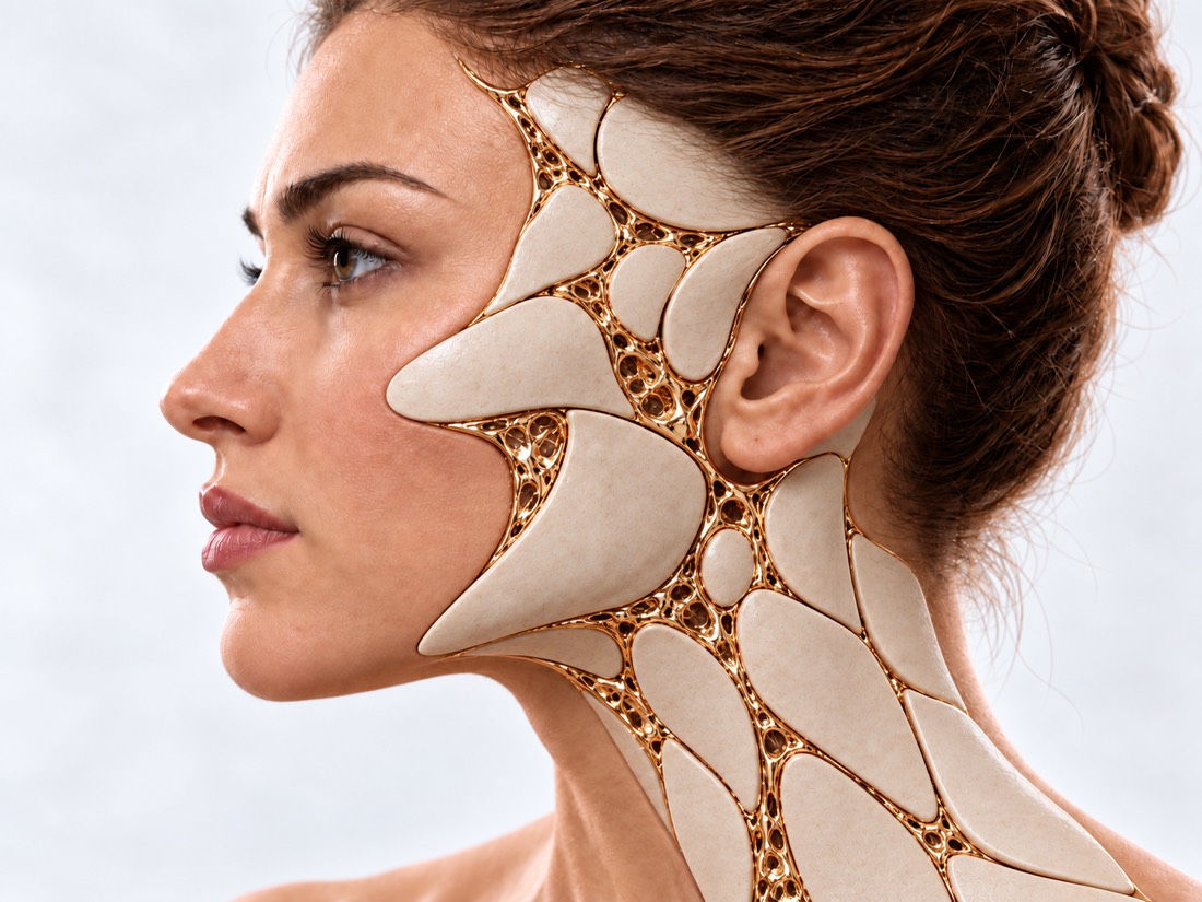
Conceptual visualization: skin and soft tissues of the faceCONCEPTUAL VISUALIZATION
What it is
Benign tumor from blood vessels; the most common is infantile, appearing in the first weeks of life.
How common
Up to 5% of babies are the most common benign tumor at this age.
How it grows
It reaches 80% of its size by 3 months, and then slowly decreases over the years.
Who treats it
Medicines are prescribed by the child's attending physician; the surgeon corrects the remnants after reduction.

What is
hemangioma

Hemangioma is a benign tumor of blood vessel cells. Infantile hemangioma occurs most often in children, and "strawberry spot" occurs in everyday life. It appears in the first weeks of life, grows for several months, and then slowly decreases. Up to 5% of babies have it; more often - girls, premature children, children with low birth weight and twins.

Specialists distinguish between vascular formations according to the classification of ISSVA - the International Society for the Study of Vascular Anomalies. She divides them into two large groups: vascular tumors, to which a hemangioma belongs, and vascular malformations — defects in the structure of vessels that a person is born with. These are different conditions with different treatments.

"Hemangioma" is often mistakenly called any vascular spot or nodule. In an analysis of 320 scientific publications, the word was used incorrectly 71.3% of the time, and patients with the incorrect name were more likely to receive inappropriate treatment. Correctly named hemangiomas were an average of 4 months old in children, and wrongly named ones were an average of 36 years old in people. Therefore, in an adult, "hemangioma" is often found to be another vascular formation, for example, a malformation.

Most hemangiomas are small, harmless, and do not require treatment. But on the face, some of them can leave persistent skin changes or interfere with seeing, breathing or eating - they should be treated in time.

This is general information. What will suit your child or you, the doctor will tell after the examination.

How does it look
hemangioma

At birth, an infantile hemangioma is usually not visible or only a faint spot is visible. It becomes noticeable in the first weeks and grows rapidly.

01

Red bulge

Superficial hemangioma is a bright red soft mass raised above the skin.

02

Bluish node

A deep hemangioma lies beneath the skin, and the skin above it is bluish or purple. Such hemangiomas usually grow later and longer than superficial hemangiomas.

03

A large spot on part of the face

Segmental hemangioma occupies the entire face and often continues to grow after 3 months.

04

Ulcer

The surface of the hemangioma disintegrates, hurts and may bleed; a scar may remain after healing.

05

It is present at birth

Congenital hemangioma is a rare variant: it is already present at birth and does not grow after it. Some such hemangiomas quickly decrease in the first months, others remain.

06

Remainders after reduction

Vascular "nets", excess skin, soft fatty-fibrous tissue or thin wrinkled skin at the site of the hemangioma.

Scars on the face

Who has it?
and where it is more risky.

Depending on the location and size, whether it is possible to simply observe or whether it is necessary to treat early.

Who more often

Girls, premature babies (born before 37 weeks), low birth weight babies and twins.

Near the eye

A hemangioma can cover the eye or press on it and impair vision. In a study of 89 children, 60% had eye complications: most often astigmatism (33%) and closed field of vision (29%). The risk is higher if the hemangioma is larger than 1 cm, deep, or on the upper eyelid.

Neoplasms of the eyelids

Nose and lip

Hemangiomas near the nose, mouth, and throat can impair function, interfere with feeding, and leave noticeable changes on the face.

Beard zone

The areas in front of the ears, the chin, the lower lip and the front surface of the neck. Such a hemangioma can be accompanied by a hemangioma in the respiratory tract: in a series of observations, 10 of 16 children had it, four of them required tracheotomy.

Large facial hemangioma

A large segmental hemangioma of the face can be combined with changes in the vessels of the brain and the aortic arch - then the doctor will refer you for an examination.

When to seek medical advice?

Most hemangiomas do not require emergency care. But a hemangioma in the "beard" area or on the neck can affect the respiratory tract, and then some signs require immediate help. Also immediately - bleeding from the ulcer, which cannot be stopped.

103 immediately if:

  • it is difficult for the child to breathe - he croaks or pulls his stomach under the ribs;
  • a high-pitched whistling sound (stridor) is heard during inhalation;
  • lips or skin are very pale, blue or gray;
  • the child cannot swallow or is drooling;
  • the child suddenly became lethargic and unresponsive as usual;
  • bleeding from a hemangioma does not stop, even if you press it tightly with a clean cloth.

Do not delay a visit to the doctor:

In the coming days: a hemangioma near the eye, on the nose, lip, in the "beard" area or large on the face; it grows quickly, is covered with an ulcer, hurts or bleeds. Such children are advised to see a doctor as early as possible - ideally before one month of age, because hemangiomas grow the fastest between 1 and 3 months. Planned: a small hemangioma that does not grow and does not interfere, as well as remnants after its reduction in an older child or adult.

As defined,
what is it and whether to treat it.

  1. Conversation and photo

    When a hemangioma was noticed, how quickly it grows; photos from the first weeks help to assess growth.

  2. Review

    The doctor determines the type—superficial, deep, segmental, or congenital—and whether it is a hemangioma at all, rather than a malformation.

  3. Examination of the eye or respiratory tract

    With a hemangioma near the eye, the child is examined by an ophthalmologist; in the "beard" area, the doctor assesses whether there is damage to the respiratory tract.

  4. Examination as needed

    If the hemangioma is deep, large, or the diagnosis is unclear, the doctor may order an ultrasound, MRI, or vascular examination.

How we treat
hemangioma

Most hemangiomas are not treated, but observed. Treatment is needed if the hemangioma threatens vision, breathing or feeding, is covered with an ulcer, or can leave noticeable changes on the face.

01

Follow-up

Small hemangiomas without risk.

The first months
A hemangioma grows fastest between 1 and 3 months, and growth is mostly complete by 5 months. Therefore, at this time, she is being watched especially carefully.
Reduction
Lasts for years: In a study of 184 untreated hemangiomas, reduction was complete in an average of 3.5 years.
02

Medicines

First line for high-risk hemangiomas.

Propranolol
The drug of choice according to the guidelines of the American Academy of Pediatrics in 2019. It is usually given orally for at least 6 months, often until the child is a year old. In a randomized trial of 460 infants, complete or near complete resolution of the hemangioma after 24 weeks was seen in 60% of infants versus 4% in the placebo group.
Security
Possible side effects include a decrease in blood sugar and blood pressure, a slower pulse, and bronchial spasm. They occurred infrequently in the study, with no significant difference from the placebo group. To prevent blood sugar from falling, propranolol is given during or immediately after feeding, and if the child does not eat well or vomits, the dose is skipped and the doctor is consulted. Treatment is prescribed and monitored by a doctor.
Repeat course
10% of children who were helped by propranolol later needed retreatment.
Timolol
For individual small, thin superficial hemangiomas - a drug that is applied to the skin.
03

Surgery and laser

Mostly for leftovers.

Leftovers
In a study of 184 untreated hemangiomas, noticeable residual changes remained in 54.9%: most often vascular "nets", fatty-fibrous tissue and thin wrinkled skin.
when
Surgery and laser are most often used for residual changes after hemangioma reduction, less often - earlier.
How they operate
Excess skin and fat-fibrous tissue are cut out, and the seam is placed in natural folds or on the border of facial areas. In a series of 107 patients, 22.4% underwent such an operation after hemangioma reduction — most often with hemangiomas of the head and neck.
04

Congenital hemangiomas

It is rapidly decreasing
This variant usually does not bother, but at the beginning it can be covered with an ulcer or bleed; decreases in the first months of life.
Does not decrease
It does not cause complications, but remains. What to do with her, the doctor decides after the examination.

How
we treat it.

Facial hemangiomas are treated at Dr. Valikhnovskyi's Institute of Surgery in the field of "Congenital Defects Surgery". The clinic is responsible for the surgical part of the treatment. Medicines such as propranolol are prescribed and monitored by the child's doctor; with a hemangioma near the eye, the child is examined by an ophthalmologist.

CLINIC SERVICE Surgery of birth defects Congenital defects of the face and body in children and adults — from the first operation to the correction of the consequences. View service

What we do for this condition

  • Microsurgical technique — magnifying optics and thin suture material help to accurately match the tissues — this is important for a less visible scar.
  • Expander — if it is necessary to close a large area, a balloon is placed under the healthy skin next to it, the skin is gradually stretched and moved to the defect site — often in several stages.
  • Lipofilling — smoothing of the soft tissues of the face with own fat.
  • Phased treatment as growth progresses — interventions are planned taking into account the child's age, and follow-up examinations continue while the child grows.

Rostislav Valikhnovskyi

Plastic surgeon, candidate of medical sciences, founder of Dr. Valikhnovskyi Institute of Surgery.

How treatment works with us

  1. Photo onlineSend a photo of the hemangioma directly and from the side and tell us when it was noticed.
  2. ConsultationOverview, location and risk assessment; if necessary, referral to an ophthalmologist or ENT doctor.
  3. PlanObservation, medicine or surgery - and when exactly.
  4. SurgeryIf necessary — removal of residual tissues with a seam in the natural lines of the face.
  5. ControlExaminations after surgery and as the child grows.

Common
questions

Short answers. What is right for you, the doctor will tell you during the consultation.

Will the hemangioma go away by itself?

Most small hemangiomas shrink without treatment, but slowly over years. More than half of untreated hemangiomas leave behind skin changes: in a study of 184 hemangiomas — 54.9%.

When to show a hemangioma to a doctor?

If it is near the eye, nose, mouth, in the "beard" area, is large, grows quickly, hurts or is covered with an ulcer - as early as possible, ideally before a month of life.

Is propranolol safe for a baby?

A Cochrane review found no difference in the rate of serious side effects compared to placebo. A decrease in blood sugar and pressure, a slower pulse, and bronchial spasm are possible, so the treatment is prescribed and monitored by a doctor.

How long does propranolol treatment last?

Usually at least 6 months, often up to the child's year, sometimes longer.

When can the remnants be removed by surgery?

Usually after the hemangioma has shrunk. Sometimes they operate earlier - the doctor decides after the examination.

I, an adult, have a "hemangioma". What could it be?

Infantile hemangioma appears in the first weeks of life. In adults, "hemangioma" is often called other vascular formations, in particular malformations, and treatment depends on the exact diagnosis.

Can a hemangioma interfere with breathing?

Maybe if it is in the respiratory tract. A hemangioma in the "beard" area is a reason to check the respiratory tract. If the child has difficulty breathing or hears whistling when breathing in, call 103.

Sources

18 sources · AAP guideline, ISSVA classification, reviews, research and NHS
showCollapse
  1. Clinical Practice Guideline for the Management of Infantile HemangiomasPediatrics, 2019doi:10.1542/peds.2018-3475 ↗
  2. Vascular Anomalies Classification: Recommendations From the International Society for the Study of Vascular AnomaliesPediatrics, 2015doi:10.1542/peds.2014-3673 ↗
  3. Interventions for infantile haemangiomas of the skinCochrane Database of Systematic Reviews, 2018doi:10.1002/14651858.CD006545.pub3 ↗
  4. Infantile haemangiomaLancet, 2017doi:10.1016/S0140-6736(16)00645-0 ↗
  5. A randomized, controlled trial of oral propranolol in infantile hemangiomaNew England Journal of Medicine, 2015doi:10.1056/NEJMoa1404710 ↗
  6. Growth characteristics of infantile hemangiomas: implications for managementPediatrics, 2008doi:10.1542/peds.2007-2767 ↗
  7. Risk Factors for Degree and Type of Sequelae After Involution of Untreated Hemangiomas of InfancyJAMA Dermatology, 2016doi:10.1001/jamadermatol.2016.2905 ↗
  8. How to Manage Disfiguring Scars in Involuted Infantile HemangiomaAdvances in Wound Care, 2019doi:10.1089/wound.2018.0847 ↗
  9. Increased risk of symptomatic hemangiomas of the airway in association with cutaneous hemangiomas in a "beard" distributionJournal of Pediatrics, 1997doi:10.1016/s0022-3476(97)70079-9 ↗
  10. Subglottic hemangioma: Understanding the association with facial segmental hemangioma in a beard distributionInternational Journal of Pediatric Otorhinolaryngology, 2018doi:10.1016/j.ijporl.2018.07.019 ↗
  11. Periocular infantile hemangiomas: Characteristics, ocular sequelae, and outcomesPediatric Dermatology, 2019doi:10.1111/pde.13925 ↗
  12. Risk factors for ocular complications in periocular infantile hemangiomasPediatric Dermatology, 2018doi:10.1111/pde.13525 ↗
  13. Evaluation of terminology for vascular anomalies in current literaturePlastic and Reconstructive Surgery, 2011doi:10.1097/PRS.0b013e3181f95b83 ↗
  14. Congenital hemangiomasSeminars in Cutaneous Medicine and Surgery, 2016doi:10.12788/j.sder.2016.045 ↗
  15. BirthmarksNHS, 2023nhs.uk/conditions/birthmarks ↗
  16. Shortness of breathNHS, 2024nhs.uk/symptoms/shortness-of-breath ↗
  17. Sore throatNHS, 2024nhs.uk/symptoms/sore-throat ↗
  18. Cuts and grazesNHS, 2026nhs.uk/conditions/cuts-and-grazes ↗

The figures on the page are from these sources.

VALIKHNOVSKI SURGERY PARTNERS

A hemangioma was noticed
in a child?

Send a photo and tell us when you spotted it. The doctor will tell you whether it is possible to observe, or whether it is better not to delay.

Call Send an enquiry
ONLINE ENQUIRY

Tell us how we can help.

Leave your contact details and briefly describe the situation. A coordinator will contact you to agree on the next step.

Preferred contact method