CONDITION / SKIN AND SCARS

Giant congenital nevus.

A giant congenital nevus is a large birthmark that a child is born with and that grows along with them. It is rare, so it is important for parents to understand what really needs to be monitored and when surgery is helpful.

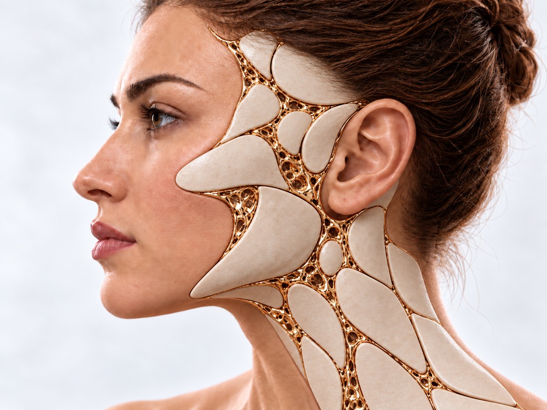
Conceptual visualization: skin with a large congenital pigmented spotCONCEPTUAL VISUALIZATION
What it is
A congenital pigment spot that will reach over 40 cm in adulthood.
How common
Large congenital nevi occur in 1 in 20,000 to 1 in 500,000 newborns.
What to monitor
Risk of melanoma and changes in the nervous system; for the latter, an MRI is done.
Who treats it
Monitored by a dermatologist and neurologist, gradually removed by a plastic surgeon.

What is
Giant nevus.

Congenital nevus — a cluster of pigment cells in the skin that is present from birth. The nevus grows proportionally with the child, so its size is assessed based on what it will be in adulthood. A nevus over 20 cm is called large, over 40 cm — giant.

Such a nevus can be brown or black, may darken over time, become raised, and grow hair, especially during adolescence. The surface can be uneven, with small nodules occurring. Nearby and throughout the body, there can be smaller nevi — these are called satellites.

Large congenital nevi are rare: from 1 in 20,000 to 1 in 500,000 newborns. Doctors monitor two things. First — the risk of melanoma, a cancer of the pigment cells. Second — neurocutaneous melanosis: pigment cells can also be in the brain or spinal cord, so some children undergo MRI. Most children do not have these complications: in a review of 14 studies (2,578 patients with large nevi), melanoma developed in 2%, and in a meta-analysis, neurological signs were present in 7.04% of patients with congenital nevi. It is important to be aware of these risks in order to notice changes in time.

Treatment of a large nevus often consists of several stages, so it is planned over years together with the parents.

This is general information. What will suit your child, the doctor will tell after the examination.

How does it look
large nevus.

The nevus has been visible since birth. The doctor describes not only its size, but also the surface, hair, nodules, and the number of satellites — this determines the examination plan.

01

Large spot

Brown or black, covering a significant part of the back, torso, limbs, or head. It can darken and become raised with age.

02

Satellites

Smaller nevi in other locations. Their number is recorded in the first year of life: it affects the examination plan, including the decision about MRI.

03

Hair and unevenness

Hair may grow on a nevus; the surface can be bumpy, and nodules may occur in the skin or beneath it.

04

Itching

The skin on a nevus may itch. Itching and pain reduce the child's quality of life, so it is worth telling the doctor about them.

05

Changes in the nevus

A nevus grows along with the child and may darken, become raised, and grow hair with age. Changes in a specific area should raise concern: a new nodule, an area that grows faster than the rest of the nevus, changes color, hurts, becomes inflamed, bleeds, or forms a crust. Report any changes to the doctor.

Melanoma of the skin of the face
06

Neurological signs

Developmental delay and seizures are signs most often reported in children with changes in the nervous system.

It depends on what
risk

A nevus arises due to excessive growth of pigment cells in the skin before birth. The risks vary in different children: they depend on the size of the nevus, satellites, and the number of nevi.

Size

In a review of 7,915 patients with congenital nevi of all sizes, melanoma developed in 1.15%. In large and giant nevi, the frequency of melanoma up to 15 years of age was 21.9 times higher than in small and medium nevi. In another review of 2,578 patients with large nevi, in 74% of melanoma cases the nevus was over 40 cm.

Satellites

In a study of 120 children, in 16 who had no satellites at birth, there were no complications. Overall, MRI or neurological changes were found in 18%, more often — with nevi over 40 cm.

Multiple nevi

In a study of 46 children who had MRI due to a nevus, pigment in the nervous system was found in 8 (17%), and all of them had more than 4 congenital nevi.

Location on the body

In a review of 7,915 patients, melanoma more often occurred when the nevus was on the torso. For changes in the nervous system, the location of the nevus, particularly along the spine, did not matter in a study of 120 children.

When to seek medical advice?

The nevus itself does not require emergency care. But some signs in a child, especially after surgery, require quick action.

103 immediately if:

  • seizures in the child;
  • the child is breathing very fast or with difficulty, grunting or pulling in the abdomen during inhalation;
  • skin, lips, or tongue have turned blue, gray, pale, or mottled;
  • The child's skin is cold or sticky to the touch;
  • a rash that does not fade when pressed;
  • a temperature of 38 °C and above in a child up to 3 months or 39 °C and above — in 3–6 months;
  • the temperature is below 36 °C, the child is cold to the touch or shivering;
  • weak, shrill or continuous crying;
  • The child has fainted, and it is difficult or impossible to wake them;
  • the child did not urinate for 12 hours;
  • Swollen lips, tongue, or throat, the child has difficulty swallowing or speaking;
  • A child from 5 years old or an adult is confused, speaks unclearly or nonsensically;
  • A child from 5 years old or an adult has a very high or very low temperature, shivers;
  • Bleeding from a wound or nevus does not stop, even though you press the wound tightly.

See a doctor without delay if:

Immediately - to the surgeon who operated, and if it does not work - to the waiting room: After surgery, pain or swelling in the area of the expander or wound increases, the suture has come apart, the wound is red or pus is coming from it. On the same day: Possible signs of fluid accumulation in the brain (hydrocephalus) — an infant's head is unusually large, the fontanelle is bulging or tense, eyes look downward, the child eats poorly, vomits, is irritable or unusually sleepy; in an older child — headache and vomiting, worse in the morning, unusual drowsiness, double or blurred vision, unsteadiness while walking. In the coming days: A new nodule or area has appeared in the nevus that is growing faster than the rest of the nevus, changing color or shape, is painful, inflamed, bleeding, or has formed a scab; the child has started to lag in development or is losing skills they previously had. If the doctor suspects melanoma, an oncologist should be consulted within 2 weeks. Planned: The child was born with a large nevus — consult a dermatologist to create a monitoring plan and discuss MRI and treatment.

How we assess
a child

  1. Examination and measurements

    The doctor measures the nevus, predicts its adult size, counts satellites, and describes the surface, hair, and nodules.

  2. Photos and monitoring

    The nevus is photographed and the images are compared year to year: this shows whether it is lightening, growing, or changing.

  3. MRI

    Of the brain and spinal cord — for children with large or multiple congenital nevi. In a study of 289 children with multiple nevi, MRI changes were found in 21%. There are no strong recommendations for MRI for everyone, so the decision is made individually.

  4. Neurologist

    If MRI shows changes or there are seizures or developmental delays. When the neurological state changes suddenly, MRI is repeated with contrast.

  5. Histology

    Nodules that raise doubt and tissue removed during surgery are examined under a microscope.

How we treat
large nevi.

There is no single plan for everyone. Doctors weigh the size and location of the nevus, the child’s age, surgical risks, and family preferences.

01

Observe or operate

The decision is made together with the parents.

Not “at any cost”
According to modern recommendations, the main focus of treatment is the appearance in the long-term perspective. The approach of "removal at any cost" is no longer used, and possible complications are weighed against expected benefits.
Does surgery reduce the risk
In a review of 7,915 patients, melanoma was more often found in nevi that were not removed or partially removed than after complete excision. At the same time, another review notes: it has not been proven that planned surgery reduces the risk of cancer, so it is performed primarily for the sake of appearance.
when
A nevus without treatment may lighten, sometimes noticeably. The benefits of early surgeries have not been proven, and anesthesia risks in infants are higher, so in some centers planned surgeries are postponed at least until the age of one.
02

How to delete

A large nevus is removed in several stages.

Expander
A balloon is injected next to healthy skin and filled over weeks, then the nevus is excised and covered with the stretched skin. In a series of 14 patients, the expanders were removed on average after 84.71 days; complications occurred in 2: wound dehiscence in one, expander infection in the other.
Several cycles
One stretch is not always enough. In a study of 55 patients, 31 had successive cycles; complications were more often associated with large expanders and large filling volumes.
Dermabrasion and laser
After dermabrasion, wounds often heal worse, leaving coarse scars, and pigmentation returns after years. Laser temporarily lightens the nevus but often leaves scars; there is no reliable long-term outcome data.
03

Years of follow-up

Skin examinations
Regularly, especially in childhood: that is when the risk of melanoma is considered highest.
Neurologist
For children with MRI changes or neurological signs — according to a separate plan.
Child support
According to a survey of parents of 235 children, quality of life was reduced by neurological problems, itching or pain, and feelings of stigma. The size of the nevus and whether it had been removed were not significant factors in this survey. Therefore, both skin care and psychological support are important.

How
we treat it.

Large and giant congenital nevi at the Doctor Valikhnovskyi Institute of Surgery are treated under the 'Surgery of Congenital Anomalies' program — step by step, with expanders, to cover the area with the patient’s own skin. Monitoring of the nevus, MRI, and neurological issues are managed by a dermatologist and a neurologist; the clinic is responsible for the surgical part — removal of the nevus and skin reconstruction.

CLINIC SERVICE Surgery of birth defects Congenital defects of the face and body in children and adults — from the first operation to the correction of the consequences. View service

What we do for this condition

  • Expander for nevus — a silicone balloon is placed under healthy skin nearby and filled with saline for several weeks; then the nevus is excised, and the area is covered with stretched skin from the patient, similar in color and texture.
  • Stepwise treatment — large nevi are removed in several stages, according to the child’s growth, taking into account their age and size.
  • Expander on the face and head — the balloon is placed on the cheek, neck, forehead, or scalp and filled for 1–3 months to move healthy skin to the site of the nevus.
  • Local and perforator flaps — on the face, the wound is covered with neighboring tissue without rough tension and without distorting the lines of the mouth or eyes.
  • Parents nearby — in the hospital, parents stay with the child; afterwards — follow-up examinations as the child grows.

Rostislav Valikhnovskyi

Plastic surgeon, candidate of medical sciences, founder of Dr. Valikhnovskyi Institute of Surgery.

How treatment works with us

  1. Photo onlineSend a photo of the nevus and satellites, as well as MRI results and doctors' conclusions, if available.
  2. ConsultationExamination of the nevus, discussion about the child's age, examination and family expectations.
  3. Plan of stagesHow many expanders, where to place them, and in what sequence to remove the nevus.
  4. OperationsPlacement of an expander, filling over weeks, removal of the nevus area and skin transfer.
  5. SupervisionMonitoring healing, histology results, and check-ups as the child grows.

Common
questions

Short answers. What is right for you, the doctor will tell you during the consultation.

Will the nevus become melanoma?

In most children — no. In a review of 2,578 patients with large nevi, melanoma developed in 2%. Among these melanomas, 74% arose in nevi over 40 cm, and 94% of patients with melanoma had satellites. Therefore, regular check-ups are important.

Why an MRI if there is a nevus on the skin?

Pigment cells can also be present in the brain or spinal cord. In a study of 289 children with multiple nevi, MRI changes were found in 21%; among those who had only pigment deposits in brain tissue, no malignant tumors developed during the observation period. Whether an MRI is needed is decided by the doctor.

When is it better to operate?

There is no specific age. The benefits of early surgeries are not proven, and the risks of anesthesia in infants are higher. Large nevi are removed in stages as the child grows, and when to start is decided by the doctor together with the parents after an examination.

Can a giant nevus be removed all at once?

Large nevi are removed in stages, often with expanders: one skin stretch is not always enough to cover the entire area. How many stages and expanders will be needed is decided by the doctor based on the size and location of the nevus.

What is an expander?

A silicone balloon that is placed under healthy skin next to the nevus and gradually filled. The skin stretches and is used to cover the area of the removed nevus. Possible complications include wound dehiscence or expander infection.

Will laser or dermabrasion help?

Both methods often leave scars, and the pigment returns. There is no reliable long-term data for the laser.

Can a nevus lighten on its own?

It can, sometimes noticeably. And sometimes it's the opposite: with age, especially during adolescence, the nevus darkens, becomes raised, and grows hair. That is why it is photographed and the images are compared.

Sources

21 sources · reviews, guidelines and research
showCollapse
  1. Care of Congenital Melanocytic Nevi in Newborns and Infants: Review and Management RecommendationsPediatrics, 2021doi:10.1542/peds.2021-051536 ↗
  2. Multidisciplinary long-term care and modern surgical treatment of congenital melanocytic nevi - recommendations by the CMN surgery networkJournal der Deutschen Dermatologischen Gesellschaft, 2019doi:10.1111/ddg.13951 ↗
  3. New recommendations for the categorization of cutaneous features of congenital melanocytic neviJournal of the American Academy of Dermatology, 2013doi:10.1016/j.jaad.2012.05.043 ↗
  4. Neurological signs, symptoms and MRI abnormalities in patients with congenital melanocytic naevi and evaluation of routine MRI-screening: systematic review and meta-analysisOrphanet Journal of Rare Diseases, 2022doi:10.1186/s13023-022-02234-8 ↗
  5. Central nervous system magnetic resonance imaging abnormalities and neurologic outcomes in pediatric patients with congenital nevi: A 10-year multi-institutional retrospective studyJournal of the American Academy of Dermatology, 2022doi:10.1016/j.jaad.2022.05.062 ↗
  6. Classification of neurological abnormalities in children with congenital melanocytic naevus syndrome identifies magnetic resonance imaging as the best predictor of clinical outcomeBritish Journal of Dermatology, 2015doi:10.1111/bjd.13898 ↗
  7. Large congenital melanocytic nevi: therapeutic management and melanoma risk: a systematic reviewJournal of the American Academy of Dermatology, 2013doi:10.1016/j.jaad.2012.09.039 ↗
  8. Melanoma in congenital melanocytic naeviBritish Journal of Dermatology, 2017doi:10.1111/bjd.15301 ↗
  9. Complications of congenital melanocytic naevi in children: analysis of 16 years' experience and clinical practiceBritish Journal of Dermatology, 2008doi:10.1111/j.1365-2133.2008.08775.x ↗
  10. Risk of melanoma in congenital melanocytic nevi of all sizes: A systematic reviewJournal of the European Academy of Dermatology and Venereology, 2023doi:10.1111/jdv.18581 ↗
  11. The role of surgery in the management of congenital melanocytic naevi in children: a perspective from Great Ormond Street HospitalJournal of Plastic, Reconstructive & Aesthetic Surgery, 2009doi:10.1016/j.bjps.2008.12.016 ↗
  12. Multiple Tissue Expansion for Giant Congenital Melanocytic NevusAnnals of Plastic Surgery, 2017doi:10.1097/SAP.0000000000001215 ↗
  13. Multifactorial analysis of the surgical outcomes of giant congenital melanocytic nevi: Single versus serial tissue expansionArchives of Plastic Surgery, 2020doi:10.5999/aps.2020.01494 ↗
  14. Predictors of Health-related Quality of Life and Psychological Adjustment in Children and Adolescents With Congenital Melanocytic Nevi: Analysis of Parent ReportsJournal of Pediatric Psychology, 2019doi:10.1093/jpepsy/jsz017 ↗
  15. Laser treatment of congenital melanocytic naevi: a systematic reviewBritish Journal of Dermatology, 2018doi:10.1111/bjd.16094 ↗
  16. When to get urgent medical help for babies and children under 5NHS, 2026nhs.uk/baby/health/when-to-get-urgent-medical-help-for-babies-and-children-under-5 ↗
  17. SepsisNHS, 2026nhs.uk/conditions/sepsis ↗
  18. BirthmarksNHS, 2023nhs.uk/conditions/birthmarks ↗
  19. MolesNHS, 2023nhs.uk/conditions/moles ↗
  20. Symptoms of hydrocephalusNHS, 2023nhs.uk/conditions/hydrocephalus/symptoms ↗
  21. Cuts and grazesNHS, 2026nhs.uk/conditions/cuts-and-grazes ↗

The figures on the page are from these sources.

VALIKHNOVSKI SURGERY PARTNERS

The child was born
with a large nevus?

Send a photo of the nevus and doctors’ reports. The surgeon will say whether surgery is needed, when it is best to plan it, and how many stages may be required.

Call Send an enquiry
ONLINE ENQUIRY

Tell us how we can help.

Leave your contact details and briefly describe the situation. A coordinator will contact you to agree on the next step.

Preferred contact method