CONDITION / CARRY

Cyst of the maxillary sinus.

Cyst of the maxillary sinus is a benign bubble with fluid in the mucous membrane of the sinus. Most often, it is found accidentally on a CT scan, and it does not require treatment; remove only the cysts that cause complaints.

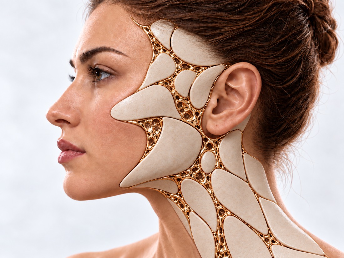
Conceptual visualization: sacral sinusesCONCEPTUAL VISUALIZATION
what is it
A benign bubble with fluid in the mucous membrane of the maxillary sinus.
How often
Depending on the examination method — in 3.6–35.6% of people; on CT of the sinuses in one study of 5166 people — in 27.6%.
Is it dangerous?
Usually no: most cysts do not cause symptoms and do not require surgery.
Who treats
ENT doctor; the operation is performed endoscopically, through the nose.

What is
sinus cyst.

A retention cyst is a fluid-filled bubble in the mucous membrane of the maxillary sinus. It occurs when the duct of the mucous gland is blocked. In the picture, it looks like a rounded shadow, usually at the bottom of the sinus.

Such cysts are very common and mostly safe. They are not as closely related to nasal complaints as they seem: in prospective studies, cyst did not correlate with either symptoms or the severity of changes on CT.

Another situation is a mucocele: a cyst that fills and expands the sinus, presses on its walls and can affect the eye. Mucocele is treated surgically.

This is general information. Whether you need treatment, the doctor will tell after the examination and CT scan.

How a
cyst manifests itself.

More often than not. Sinus cysts are among the most frequent incidental findings on a CT scan of the head taken for another reason.

01

Without symptoms

Most of the cysts a person does not feel and learns about them from the picture.

02

Heaviness in the cheek

A large cyst or mucocele may cause pressure or pain on one side of the face.

03

Congestion and discharge

If the cyst interferes with the outflow from the sinus.

Chronic sinusitis
04

Cheek swelling or numbness

More typical for mucocele, which expands the sinus.

05

Displacement of the eye

A rare sign of a mucocele pressing on the eye socket requires urgent examination.

Why does a
cyst appear.

Why retention cysts are formed is not fully understood.

Anatomy of the nose

In a study of 5166 CT scans, 88.7% of cysts were accompanied by at least one feature of the structure of the nose or sinuses.

Distortion of the septum

Inflammation

The following factors are described for mucocele: chronic sinusitis, previous operations, allergic rhinitis, nasal trauma; in 56% the cause was not found.

Natural course

Cysts can change: decrease in about 30%, do not change in 50-60%, grow in 8-20%.

When to seek urgent medical care?

A retention cyst does not require emergency care. An urgent examination is required if there are signs of pressure on the eye - they can be caused by a mucocele.

103 immediately if:

  • sudden deterioration of vision, double vision or displacement of the eye;
  • swelling and redness of the eyelids with temperature.

See an ENT doctor in the coming days if:

Pain, swelling or numbness of the cheek on one side is increasing, or bleeding has appeared. If the cyst was just found on the image, make an appointment for a scheduled consultation to assess whether something needs to be done.

How to define
tactics.

  1. Examination and endoscopy

    The doctor evaluates whether there are complaints that could really be from a cyst.

  2. CT scan of the sinuses

    Shows the size of the cyst and the condition of the sinus opening; distinguishes a cyst from a mucocele.

  3. Before implantation

    If a sinus floor lift is planned for dental implants, the dentist and ENT evaluate the cyst together.

  4. Observation

    If the cyst does not interfere, repeat the picture if necessary.

How to treat a
sinus cyst.

Cysts of the maxillary sinus are mostly harmless formations without symptoms that usually do not require surgery. Remove those that cause complaints.

01

Without surgery

Observation
If the cyst does not cause symptoms, it is enough to know about it; medicines do not dissolve it.
02

Endoscopic surgery

Through the nose, without incisions on the face.

Micropharyngotomy
The surgeon expands the sinus opening and removes the cyst under the control of the endoscope.
The main thing is ventilation
In a randomized trial of 80 patients with large, complaining cysts, outcome was independent of whether the cyst itself was removed: it was important to restore sinus ventilation and drainage.
Mucocele
Treated endoscopically. In a study of 36 patients, symptoms disappeared in all of them, and repeated operations were not required.
03

Cyst and dental implants

Before lifting the sinus floor
A review of 19 studies (182 patients) found no difference in implant outcomes between whether the cyst was removed or not. Decisions are made individually.

How do we
treat it.

Cysts of the maxillary sinuses, which cause complaints, are removed at the Institute of Surgery of Dr. Valikhnovsky in the direction of "New Nose" - endoscopically, through the nostrils.

DIRECTION TO THE CLINIC A new nose Shape, support and breathing of the nose: septum, concha, sinuses, consequences of injuries, failed operations and removal of tumors. Go to direction

What do we do in this condition

  • Endoscopic microhygrotomy — expansion of the sinus opening and removal of the cyst.
  • Revision of the ostiomeatal complex — restoration of sinus outflow tracts.

Rostislav Valikhnovsky

Plastic surgeon, candidate of medical sciences, founder of Dr. Valikhnovsky Institute of Surgery.

How is the treatment in us

  1. ConsultationTalking about complaints, nasal endoscopy, looking at your CT scan.
  2. The solutionIf the cyst does not interfere, we will explain why it is not necessary to operate.
  3. PlanIf the cyst gives complaints - the extent of endoscopic surgery.
  4. OperationEndoscopically, through the nostrils.
  5. RecoverySwelling in the nose passes gradually; the doctor will explain what is possible and what should be avoided after the operation.

Frequently asked
questions.

Short answers. What is right for you, the doctor will tell you during the consultation.

Is it necessary to remove a maxillary sinus cyst?

No. Most cysts do not cause symptoms and do not require surgery. Those causing complaints or mucocele are removed.

Can the cyst dissolve by itself?

Yes, sometimes: approximately 30% of cysts shrink, 50–60% do not change, and 8–20% grow.

Is it possible to cure a cyst with drops?

No, the medicine does not dissolve the cyst. But they can reduce the accompanying swelling of the mucous membrane.

Is the maxillary sinus cyst dangerous?

A retention cyst is a harmless formation. Mucocele, which can press on the eye, is another matter; in the case of deterioration of vision, an urgent examination is required.

Does the cyst interfere with dental implantation?

In a review of 19 studies, the results of a sinus floor lift did not differ depending on whether the cyst was removed or not. The decision is made by the dentist and ENT together.

Can a mucocele affect vision?

Rarely, but yes: sinus mucocele can press on the eye socket. In a meta-analysis of 207 patients with low vision after endoscopic surgery, vision improved in 71.5%, and a better outcome was associated with surgery in the first 6 days.

Can the cyst return after surgery?

This is possible. Therefore, during the operation, it is important not only to remove the cyst, but also to restore the outflow from the sinus.

Sources

9 sources · reviews, guidelines and studies
ShowCollapse
  1. Cysts of the maxillary sinus: a literature reviewInternational Forum of Allergy & Rhinology, 2013doi:10.1002/alr.21177 ↗
  2. Symptomatic maxillary sinus retention cysts: should they be removed?Laryngoscope, 2010doi:10.1002/lary.21040 ↗
  3. Are mucous retention cysts and pseudocysts in the maxillary sinus a risk factor for dental implants? A systematic reviewMedicina Oral, Patología Oral y Cirugía Bucal, 2021doi:10.4317/medoral.24155 ↗
  4. A systematic review on incidental findings in cone beam computed tomography (CBCT) scansDentomaxillofacial Radiology, 2019doi:10.1259/dmfr.20180396 ↗
  5. Paranasal Sinus Anatomic Variations Accompanying Maxillary Sinus Retention Cysts: A Radiological AnalysisTurkish Archives of Otorhinolaryngology, 2017doi:10.5152/tao.2017.2759 ↗
  6. Correlation of quantitative MR imaging findings with symptoms in patients with incidentally detected inflammatory sinonasal diseaseDiagnostic and Interventional Imaging, 2018doi:10.1016/j.diii.2017.05.012 ↗
  7. Maxillary sinus mucocele: predisposing factors, clinical presentations, and treatmentOral and Maxillofacial Surgery, 2017doi:10.1007/s10006-016-0599-5 ↗
  8. Endoscopic Management of Paranasal Sinus Mucoceles: Meta-analysis of Visual OutcomesOtolaryngology–Head and Neck Surgery, 2017doi:10.1177/0194599817717674 ↗
  9. Immediately or delayed sinus augmentation after pseudocyst removal: A randomized trialClinical Implant Dentistry and Related Research, 2023doi:10.1111/cid.13225 ↗

The figures on the page are from these sources.

VALIKHNOVSKI SURGERY PARTNERS

Did you find a cyst
on the picture?

Send CT and describe if there are any complaints. The doctor will tell if something needs to be done - or if it is enough to observe.

to call Send an appeal