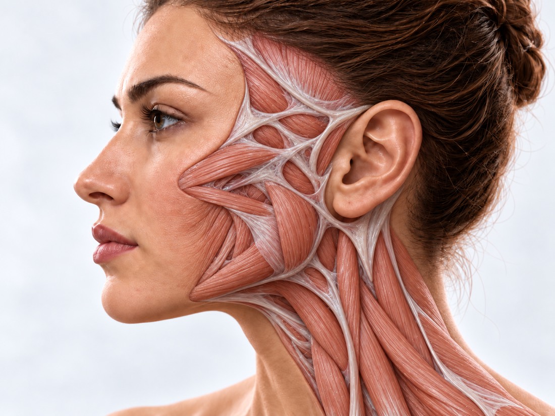
CONDITION / FACE

Skull bone defect: cranioplasty.

Cranioplasty is an operation that closes a skull bone defect after trauma, trepanation or removal of part of the bone: with one's own bone or an implant. It is planned together with a neurosurgeon to protect the brain again and restore the shape of the head.

Conceptual visualization: restoration of the cranial vault boneCONCEPTUAL VISUALIZATION
What it is
An opening or depression in the skull bone after trauma, trepanation, infection, or removal of a tumor.
What to close
Own bone or implant: titanium, PEEK, hydroxyapatite, acrylic plastic (PMMA).
why
Protect the brain, restore the shape of the head; it can also help with recovery from injury.
Who treats it
The skull bone is restored together with a neurosurgeon.

What is
skull bone defect.

Sometimes part of the skull bone has to be removed. This is done in case of a severe brain injury, when it swells and lacks space, as well as in case of a stroke, tumor or infection. This operation is called a decompressive craniectomy, and it is followed by a cranioplasty.

A bone defect remains even after injuries, infections, removal of tumors or due to congenital changes. At this point, the brain is covered only by membranes and skin.

Cranioplasty restores the bone defect. Its purpose is to restore the shape of the head, improve the movement of cerebrospinal fluid and protect the brain again. This can facilitate rehabilitation after an injury.

The skull bone is restored together with a neurosurgeon. There is a separate page about fresh fractures of the forehead called "Fractures of the frontal bone", and about scars on the forehead - "Cicatrical deformity of the forehead".

This is general information. The doctor will tell you what will suit you after the examination.

How
skull defect presents.

Some of the changes are visible and can be felt by hand. Others are in a state of well-being, and it is easy to write them off as the effects of trauma.

01

A depression or soft spot

At the site of the removed bone, there is no support under the skin: the skin sinks or, on the contrary, protrudes.

02

Inflamed skin

Months after a craniectomy, the skin over the defect may thicken deeply. Doctors call it the "inflamed flap" syndrome.

03

Health worsened

Weeks or months after craniectomy, the arm or leg gradually weakens, it becomes harder to speak, think, and drowsiness appears. This is how trepanned syndrome manifests itself — a rare complication in which the condition improves after cranioplasty.

Why does it stay?
bone defect.

Often the defect is a consequence of a life-saving operation. It can also be left behind by trauma, infection, or tumor.

Craniectomy after trauma

In case of a severe brain injury, the bone is removed so that the brain has enough space during swelling. They return it or replace it later.

Stroke, tumor, infection

The same operation is performed for severe stroke, tumors and infections. In a review of 56 patients with trepanned syndrome, trauma was the reason for craniectomy in 43%, tumor removal in 23%.

The bone dissolved

Own bone, which was returned to its place, can gradually dissolve: according to a meta-analysis of 791 such operations - in 20%. This happens more often after an injury.

Infection

Infection is the most common complication of cranioplasty: in a meta-analysis, it occurred in 8% of cases (463 of 4,667). Infected bone or an implant sometimes has to be removed.

When to seek medical advice?

A long-standing defect of the skull usually does not require emergency care - cranioplasty is planned. But without the bone, the brain in this place is less protected. According to the NHS, call 103 if you have any of the following symptoms (don't drive yourself to hospital):

103 immediately if:

  • after a blow to the head, a person faints and does not regain consciousness;
  • after a blow to the head, he cannot help but fall asleep, his eyes close by themselves;
  • convulsions began after a blow to the head;
  • vision or hearing worsened after a blow to the head;
  • after a blow - a bruise around the eye, although the eye was not hit;
  • after a blow, a clear watery liquid flows from the nose or ear;
  • after a blow - bleeding from the ear or a bruise behind the ear;
  • after a blow - new numbness or weakness in any part of the body;
  • gait, balance, understanding, speech or writing were impaired after the impact;
  • after the blow, the behavior changed: irritability, inattention, indifference to everything;
  • falling from a height of more than 1 meter or 5 steps;
  • a blow to the head at high speed, such as in a car accident;
  • a wound on the head with something stuck in it, or a new dent on the head;
  • in a child up to 1 year old, after a blow, there is a bruise, swelling or a large wound on the head;
  • sudden squinting of the face, weakness of an arm or leg, or slurred speech - even if it has passed;
  • very strong headache;
  • confusion of consciousness, the person speaks unintelligibly;
  • the neck is stiff and it hurts to look at the light;
  • very high or very low temperature, severe chills;
  • a rash that does not fade if a glass is pressed to it;
  • convulsive attack for the first time in life;
  • convulsions last more than 5 minutes;
  • after an attack, a person does not regain consciousness or a new attack begins;
  • 3 or more seizures per day.

Do not delay:

Immediately - to the surgeon who operated, and if it does not work - to the reception department: after cranioplasty, pain or swelling in the area of the operation increases, fluid oozes from the wound, the wound splits, or the implant shows through the skin. On the same day - for medical assistance after a recent blow to the head, if you have vomited or feel dizzy, take blood-thinning medication, or were intoxicated at the time of the injury. On the same day, to the surgeon: after surgery, a temperature of 38 °C and above, redness or pus in the wound area, if there are no signs from the list for 103. On the same day, to the neurosurgeon: weeks or months after a craniectomy, gradually, day by day, an arm or leg becomes weaker, it becomes harder to speak or think, sleepiness, anxiety, or depression appear, and the skin over the defect sags. Planned: an old defect, a depression on the head or forehead, an uneven contour after a previous operation - make an appointment for a consultation.

As defined,
what to cover

  1. Conversation and extracts

    Why was the bone removed when it was, was the own bone preserved, were there infections and repeated operations.

  2. CT scan of the head

    Shows the size, shape and edges of the defect. After her, a 3D model of the skull is built for the planning and manufacture of the implant.

  3. Review of a neurosurgeon

    The condition of the brain and whether there is hydrocephalus — accumulation of cerebrospinal fluid; this is taken into account when planning the terms of the operation.

  4. The skin over the defect

    The scars and the thickness of the skin are evaluated: is it enough to reliably cover the bone or the implant.

  5. Selection of material

    The material is chosen for each defect separately and the advantages and risks of each option are explained.

How to restore
skull bone

There are few high-quality studies, so there are no evidence-based guidelines — there is an international consensus on cranioplasty after trauma. There is still no common opinion about which material and which operation period is better, so the plan is drawn up individually.

01

Own bone

If it was preserved after craniectomy.

advantage
The wound over the own bone diverges less often than over implants, in particular titanium ones (meta-analysis of 30 studies).
Absorption
Own bone can be resorbed — in 15.1% (meta-analysis of 30 studies) and in 20% (another meta-analysis, 791 operations). Therefore, there are more repeat operations after it than after synthetic implants.
After an injury
If the bone was removed due to trauma, the risk of resorption and reoperation is higher. The authors of the meta-analysis suggest that an implant in such patients may partially reduce this risk.
02

Implants

Standard or individual - according to the 3D model of the skull.

Materials
Titanium, PEEK (a strong lightweight plastic), hydroxyapatite (the mineral that makes up bone), PMMA acrylic plastic.
Repeat operations
In a network meta-analysis of 31 studies, reoperation was required in 14% of cases: after titanium in 13%, after PEEK in 5% (157 cases). The authors emphasize that the optimal material has yet to be determined.
Data are contradictory
In another review of 53 studies (3,591 adults), there were more local complications (17.19%) after PEEK and more infections (7.95%) after PMMA.
Infections
In a review of 3,260 individual implants, infections occurred in 4.2–14.9%, depending on the material. But the research lasted for different times, so it is not possible to directly compare the materials.
03

Terms and risks

When to operate
In a meta-analysis of 38 studies (4,703 patients), early cranioplasty (up to 86 days after craniectomy) was associated with better functional outcomes. The overall rate of complications did not differ, but hydrocephalus occurred more frequently after early surgery.
Convulsions
Seizures occur after cranioplasty: after statistical correction — 43 cases per 1,000 person-years. After early surgery (up to 6 months) — more often.
The skin
If the skin above the defect is missing, it is restored with flaps. In a meta-analysis of 440 cases of free flaps on the scalp with a mesh implant, the flap did not heal in 7%, complications were in 21%.
Quality of life
In a review of 25 studies, physical and social functioning, appearance, and overall quality of life improved after cranioplasty.

How
we treat it.

Bone defects of the skull at Dr. Valikhnovskyi's Institute of Surgery are restored in the "New Face" direction, where the forehead and skull vault are a separate zone of reconstruction. The terms and scope of the operation are agreed with the neurosurgeon who is treating the patient.

CLINIC SERVICE A new face Form, symmetry, facial expressions and functions after injuries, wounds, burns, removal of tumors and congenital changes. View service

What we do for this condition

  • Forehead and cranial vault — bone defects and the contour of the forehead are restored with one's own bone or an implant.
  • Individual implants — from medical titanium or PEEK, made according to a 3D model that repeats the patient's anatomy.
  • Mirror CT modeling — the intact half is mirrored to reproduce a symmetrical contour.
  • Local flaps and expander — if there is a lack of healthy skin, it is moved from nearby areas or pre-stretched with an expander.
  • Free microsurgical flaps — large defects of soft tissues are closed with tissues from other parts of the body, stitching vessels together under a microscope.
  • Lipofilling — own fat provides the contour of soft tissues after reconstruction.

Rostislav Valikhnovskyi

Plastic surgeon, candidate of medical sciences, founder of Dr. Valikhnovskyi Institute of Surgery.

How treatment works with us

  1. Pictures onlineSend a CT scan of the head, statements about operations and photos - the doctor will pre-evaluate the situation.
  2. ConsultationInspection of the defect, skin and scars; if necessary, a new CT scan and examination by a neurosurgeon.
  3. PlanMaterial, terms, whether skin preparation is required; what every team specialist does.
  4. SurgeryClosing the defect with one's own bone or an implant, if necessary, together with skin plastic surgery.
  5. Follow-upFollow-up examinations and CT scans: Some complications, such as bone resorption, appear later.

Common
questions

Short answers. What is right for you, the doctor will tell you during the consultation.

Is it necessary to close the skull defect?

The decision is made together with the neurosurgeon. Cranioplasty re-protects the brain, reshapes the head, and can aid recovery from injury.

When can cranioplasty be done after trepanation?

There is no single term. In a meta-analysis of 4,703 patients, surgery before 86 days was associated with better recovery, but hydrocephalus occurred more frequently thereafter. The term is determined by a neurosurgeon.

What is better - own bone or an implant?

There is no one-size-fits-all material. The own bone can be resorbed, a wound often spreads over the implants. The material is selected for each defect separately and the risks are explained.

What is trepanation syndrome?

A rare complication after craniectomy: movements, speech, and thinking weaken after weeks or months. In a review of studies, after cranioplasty, the condition improved within a few days on average. If you notice such changes, see a neurosurgeon the same day.

Can there be convulsions after surgery?

They can In two randomized trials, prophylactic anticonvulsant drugs reduced the number of seizures by nearly 80%. Whether you need them, only a doctor decides.

Are cranioplasty performed on children?

Yes. In a review of 864 such operations in children, own bone was used in 56% of cases. Complications in children occur quite often - both with their own bone and with implants.

What to do if you hit your head in a place without a bone?

If there is even one sign from the list for 103, call an ambulance. If there was vomiting or dizziness, get medical help the same day. Do not get behind the wheel.

Sources

19 sources · consensus, systematic reviews and meta-analyses, NHS
showCollapse
  1. Consensus statement from the international consensus meeting on post-traumatic cranioplastyActa Neurochirurgica, 2021doi:10.1007/s00701-020-04663-5 ↗
  2. Complications of Cranioplasty in Relation to Material: Systematic Review, Network Meta-Analysis and Meta-RegressionNeurosurgery, 2021doi:10.1093/neuros/nyab180 ↗
  3. Complications of cranioplasty following decompressive craniectomy for traumatic brain injury: systematic review and meta-analysisActa Neurochirurgica, 2021doi:10.1007/s00701-021-04809-z ↗
  4. Cranioplasty Timing After Decompressive Craniectomy: A Meta-Analysis of 4703 PatientsNeurosurgery, 2026doi:10.1227/neu.0000000000003978 ↗
  5. The incidence and treatment of seizures after cranioplasty: a systematic review and meta-analysisBritish Journal of Neurosurgery, 2018doi:10.1080/02688697.2018.1481197 ↗
  6. Alloplastic Cranioplasty Reconstruction: A Systematic Review Comparing Outcomes With Titanium Mesh, Polymethyl Methacrylate, Polyether Ether Ketone, and Norian Implants in 3591 Adult PatientsAnnals of Plastic Surgery, 2019doi:10.1097/SAP.0000000000001801 ↗
  7. Health-related quality of life following cranioplasty - a systematic reviewBritish Journal of Neurosurgery, 2025doi:10.1080/02688697.2023.2202244 ↗
  8. Syndrome of the Trephined: A Systematic ReviewNeurosurgery, 2016doi:10.1227/NEU.0000000000001366 ↗
  9. Systematic Review of Syndrome of the Trephined and Reconstructive ImplicationsJournal of Craniofacial Surgery, 2022doi:10.1097/SCS.0000000000008724 ↗
  10. Infection rates following custom-made cranioplasty using heterologous materials: insights from a systematic review on 3260 patients with a focus on follow-up lengthNeurosurgical Review, 2025doi:10.1007/s10143-025-03818-1 ↗
  11. Outcomes of Mesh Cranioplasty in Scalp-Free Tissue Reconstruction: A Systematic Review and Meta-analysisOtolaryngology–Head and Neck Surgery, 2023doi:10.1002/ohn.199 ↗
  12. Cranioplasty after craniectomy in pediatric patients - a systematic reviewChild’s Nervous System, 2019doi:10.1007/s00381-018-4025-1 ↗
  13. Autologous Cranioplasty is Associated with Increased Reoperation Rate: A Systematic Review and Meta-AnalysisWorld Neurosurgery, 2018doi:10.1016/j.wneu.2018.05.009 ↗
  14. Cranioplasty Following Decompressive CraniectomyFrontiers in Neurology, 2020doi:10.3389/fneur.2019.01357 ↗
  15. Complications and cosmetic outcomes of materials used in cranioplasty following decompressive craniectomy - a systematic review, pairwise meta-analysis, and network meta-analysisActa Neurochirurgica, 2022doi:10.1007/s00701-022-05251-5 ↗
  16. Head injury and concussionNHS, 2025nhs.uk/conditions/head-injury-and-concussion ↗
  17. Symptoms of a strokeNHS, 2024nhs.uk/conditions/stroke/symptoms ↗
  18. MeningitisNHS, 2026nhs.uk/conditions/meningitis ↗
  19. EpilepsyNHS, 2025nhs.uk/conditions/epilepsy ↗

The figures on the page are from these sources.

VALIKHNOVSKI SURGERY PARTNERS

A defect remained
skull bones?

Send head CT and surgery reports. The doctor will tell if the defect can be closed, with what and when.

Call Send an enquiry
ONLINE ENQUIRY

Tell us how we can help.

Leave your contact details and briefly describe the situation. A coordinator will contact you to agree on the next step.

Preferred contact method