CONDITION / FACE

Craniosynostosis in a child.

Craniosynostosis is a premature growth of one or more skull seams in a child, due to which the head grows in an irregular shape. It is usually treated surgically, and in the first months of life it can be done minimally traumatically — endoscopically.

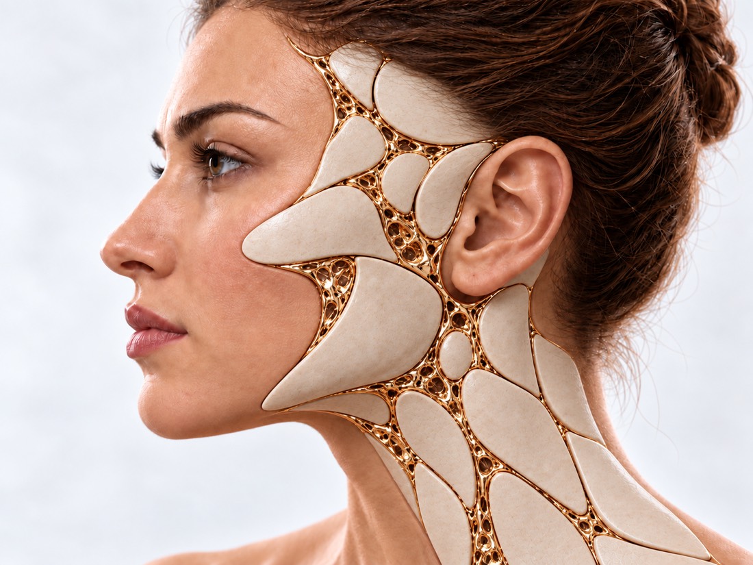
Conceptual visualization: reconstruction of the skull and face in a childCONCEPTUAL VISUALIZATION
What it is
The suture of the skull has grown earlier than it should, and the head is growing in an irregular shape.
How common
About 6 in 10,000 newborns.
What's different
Flat head from lying down (positional plagiocephaly) occurs much more often and does not require surgery.
How we treat
Surgically: in the first months — endoscopically with a helmet, later — open surgery.

What is
craniosynostosis

A baby's skull consists of several bones, between which there are flexible seams. They allow the head to grow with the brain. With craniosynostosis, one or more sutures grow prematurely: the skull stops growing in this place, and nearby grows more than necessary. This is how the shape of the head changes.

One seam or several may grow; among isolated forms, the sagittal seam, which runs along the crown, grows most often. In approximately 75% of children, craniosynostosis is isolated, that is, not associated with a syndrome. The rest are syndromic forms, when other organs are also changed.

Left untreated, craniosynostosis can cause facial deformity, increased intracranial pressure, developmental problems, and psychological difficulties. Therefore, it is important to distinguish it from a safe flat head in time.

This is general information. What will suit your child, the doctor will tell after the examination.

How to notice
craniosynostosis

The shape of the head depends on which seam has grown. The mild form is sometimes noticed only when the child grows up.

01

Long narrow head

The head is elongated like a rugby ball - this is how the fusion of the sagittal suture looks.

02

Triangular forehead

The forehead is pointed, triangular in shape — the fusion of the metopic seam in the middle of the forehead.

03

The forehead is flattened on one side

Flattening of the forehead on one or both sides - fusion of the coronal suture.

04

The back of the head is tilted

The back of the head is flattened on one side - the fusion of the lambda-like suture. It looks like a flat head from lying down, so a doctor's examination is needed.

05

Timyachko closed early

The soft crown on the top of the head disappeared within a year.

06

Signs of pressure

Repeated vomiting, unusual drowsiness, tense or bulging crown of the head are possible signs of increased intracranial pressure. They do not expect a scheduled consultation, and some of them are a reason to call 103 (see "When to contact").

Why seams?
grow early.

The exact cause is often unknown, but hereditary factors are thought to play an important role. This is a malformation of the skull, not a flat head from lying down.

Isolated form

The most common variant is found in about 75% of children: there are no other defects, and the reasons are believed to be several.

Heredity

In a survey of families of 660 patients with isolated craniosynostosis, it occurred in 7.5% of their brothers and sisters, and in 2.3% of their parents.

Syndromes

The most common are Apert, Crouzon, Pfeiffer, Müncke and Setre-Chotzen syndromes. Breathing, vision, hearing, heart and bones can be affected.

Increased pressure

Even when a single suture grew, in a summary of 17 studies, elevated intracranial pressure was found in approximately 39% of children, although the estimates depended strongly on the method of measurement.

When to seek urgent medical care?

The change in the shape of the head itself does not require emergency care. But signs of increased intracranial pressure require a doctor's examination on the same day, and some of them - a call to 103.

103 immediately if:

  • the child is difficult to wake up or is much sleepier than usual;
  • the child is lethargic, does not react as usual;
  • the crown of the head explodes or becomes tense;
  • convulsions;
  • vomit green or yellow-green, with blood or similar to coffee grounds;
  • sudden severe headache in an older child.

Other levels of urgency:

On the same day: vomiting is repeated (in a child with craniosynostosis, this may be a sign of increased pressure in the skull), the child does not retain liquid or refuses to eat - show him to the doctor on the same day. In the coming weeks: if you notice an unusual shape of the baby's head - do not delay the consultation: endoscopic surgery can be performed only in the first months of life. Planned: if the child is older, and the shape of the head, vision, hearing, speech or snoring are a concern.

How to distinguish
from a flat head.

  1. Survey and measurement

    The doctor examines the head from above, in front and behind, and measures it. Usually, it is the examination that allows you to distinguish a flat head from lying down.

  2. Flat head from lying down

    Positional plagiocephaly is a safe change in shape that occurs when a child lies on one side for a long time. It does not affect the brain and often improves within 1-2 years.

  3. Pictures

    If the examination does not give a definite answer, ultrasound or CT scan with low radiation is performed. A CT scan is not necessary to diagnose a flat head, but it helps rule out craniosynostosis.

  4. Review of other specialists

    Vision, hearing and development are checked, especially in the syndromic form.

How we treat
craniosynostosis

Surgery is needed to correct the shape of the head and give the brain room to grow. Some mild cases are only observed. The method depends on which seam has grown, on the symptoms and the age of the child.

01

Endoscopic surgery

Usually in the first months of life.

How do they do it?
Through small incisions with the help of an endoscope, the fused seam is released. After the operation, the child wears a corrective helmet for several months.
age
In a summary of 25 studies (1606 children with sagittal craniosynostosis), the average age at endoscopic surgery was 3.3 months.
Recovery
According to a meta-analysis of 34 studies (11,554 patients), after endoscopy there are fewer blood transfusions, the hospital stay is shorter by about 2.8 days, and there are 62% fewer reoperations. The long-term shape of the head is comparable to open surgery.
02

Open operation

Reconstruction of the skull vault
Changed areas of the skull are removed, modeled and fixed in the correct position. It is performed on older children when the age for endoscopy has passed.
3D planning
A digital model of the child's skull allows you to rehearse the operation in advance and print templates. According to a review of studies, with sagittal craniosynostosis, it shortened the operation by about an hour; benefits with regard to complications have not been proven.
03

After surgery

Reviews as you grow
At first — every few weeks, later less often. Vision and hearing are checked; if necessary, classes with a speech therapist and dental treatment.
Repeat operations
Some children need repeated operations in their older or teenage years.

How
we treat it.

Craniosynostosis is treated at Dr. Valikhnovskyi's Institute of Surgery in the field of "Surgery of congenital defects". Operations on the skull are planned in advance, if necessary - with 3D modeling. Sight, hearing and development after the operation are monitored by specialist children's doctors.

CLINIC SERVICE Surgery of birth defects Congenital defects of the face and body in children and adults — from the first operation to the correction of the consequences. View service

What we do for this condition

  • Endoscopic treatment of craniosynostosis — in babies up to 3–4 months — through small incisions, then a corrective helmet for several months.
  • Open skull vault reconstruction — later, when the age for endoscopy has passed.
  • 3D modeling and virtual surgery — a digital copy of the child's skull according to CT data and "virtual surgery" in advance.
  • Individual templates and stereolithographic models — printed saw guides and a skull model on which the plates are bent before surgery.
  • Distraction osteogenesis — in syndromic forms, such as Crouzon's syndrome, when facial bones are underdeveloped.

Rostislav Valikhnovskyi

Plastic surgeon, candidate of medical sciences, founder of Dr. Valikhnovskyi Institute of Surgery.

How treatment works with us

  1. Photo onlineSend a photo of the child's head from the top, front, back and side - the doctor will pre-evaluate the shape.
  2. ConsultationReview, measure and answer whether it's craniosynostosis or flat head from recumbency.
  3. Survey and planImaging as needed, 3D planning and choice between endoscopic and open surgery.
  4. SurgeryEndoscopic or open - depending on age and shape; after endoscopy - selection of a helmet.
  5. Follow-upControl of the shape of the head, wearing a helmet and the development of the child as it grows.

Common
questions

Short answers. What is right for you, the doctor will tell you during the consultation.

How to distinguish craniosynostosis from a flat head?

Flat head from lying down occurs much more often, does not affect the brain and does not require surgery. With craniosynostosis, the shape of the head is different: a long, narrow head, a triangular forehead, or a slanted forehead. A doctor's examination helps to distinguish, sometimes pictures.

Will a helmet help without surgery?

With a flat head from lying down, a helmet can speed up correction, especially if there is a pronounced deformity and early onset. With craniosynostosis, a helmet is worn after endoscopic surgery — it directs the growth of the head to the correct shape.

At what age is it better to operate?

Endoscopic surgery is performed in the first months: with the most frequent, sagittal form, the average age in the summary of studies is 3.3 months. Later - open reconstruction. Therefore, consultation should not be delayed.

Is it possible not to operate?

Some mild cases are only observed. But without treatment, craniosynostosis can cause increased pressure in the skull, facial deformity, and developmental problems, so decisions are made after an examination.

Will there be a scar?

There is a scar on the skull after surgery, but it is usually hidden by hair. After endoscopy, the incisions are small.

Will the next child have craniosynostosis?

The risk is higher than average: in a family survey of 660 patients with isolated craniosynostosis, it occurred in their siblings in 7.5%. Your doctor will discuss the risk to your family.

Does craniosynostosis affect development?

Maybe, especially if there is increased pressure in the skull. Many children have no long-term problems after treatment, but their vision, hearing and development are checked regularly.

Sources

19 sources · reviews, guidelines and research
showCollapse
  1. Global Epidemiology of Craniosynostosis: A Systematic Review and Meta-AnalysisWorld Neurosurgery, 2022doi:10.1016/j.wneu.2022.05.093 ↗
  2. Familial incidence and associated symptoms in a population of individuals with nonsyndromic craniosynostosisGenetics in Medicine, 2014doi:10.1038/gim.2013.134 ↗
  3. Prevention and management of positional skull deformities in infantsPediatrics, 2011doi:10.1542/peds.2011-2220 ↗
  4. Guidelines: Congress of Neurological Surgeons Systematic Review and Evidence-Based Guideline on the Management of Patients With Positional Plagiocephaly: The Role of RepositioningNeurosurgery, 2016doi:10.1227/NEU.0000000000001428 ↗
  5. Guidelines: Congress of Neurological Surgeons Systematic Review and Evidence-Based Guideline for the Diagnosis of Patients With Positional Plagiocephaly: The Role of ImagingNeurosurgery, 2016doi:10.1227/NEU.0000000000001427 ↗
  6. Guidelines: Congress of Neurological Surgeons Systematic Review and Evidence-Based Guideline on the Role of Cranial Molding Orthosis (Helmet) Therapy for Patients With Positional PlagiocephalyNeurosurgery, 2016doi:10.1227/NEU.0000000000001430 ↗
  7. Methodological determinants and prevalence of elevated intracranial pressure in isolated single-suture craniosynostosis: a systematic review and meta-analysisChild’s Nervous System, 2026doi:10.1007/s00381-026-07418-2 ↗
  8. Craniosynostosis: Imaging and Surgical ImplicationsRadiographics, 2026doi:10.1148/rg.250081 ↗
  9. Endoscopic versus open approach in craniosynostosis repair: a systematic review and meta-analysis of perioperative outcomesChild’s Nervous System, 2018doi:10.1007/s00381-018-3852-4 ↗
  10. Evaluating Surgical Techniques for Craniosynostosis: A Systematic Review and Meta-Analysis on Endoscopic Approach Versus Open SurgeryNeurosurgery, 2025doi:10.1227/neu.0000000000003502 ↗
  11. Open versus endoscopic surgery with helmet molding therapy in non-syndromic patients with craniosynostosis: an updated systematic review and meta-analysis of clinical outcomes and treatment-related costsChild’s Nervous System, 2024doi:10.1007/s00381-024-06692-2 ↗
  12. Shaping futures: how surgical timing influences outcomes in endoscopic sagittal craniosynostosis repairChild’s Nervous System, 2025doi:10.1007/s00381-025-06834-0 ↗
  13. The impact of virtual surgical planning on surgical outcomes in craniosynostosis surgery: a systematic review and meta-analysisNeurosurgical Review, 2025doi:10.1007/s10143-025-03973-5 ↗
  14. Isolated Sagittal Craniosynostosis: A Comprehensive ReviewDiagnostics, 2024doi:10.3390/diagnostics14040435 ↗
  15. Syndromic Craniosynostosis: A Comprehensive ReviewCureus, 2023doi:10.7759/cureus.50448 ↗
  16. CraniosynostosisNHS, 2023nhs.uk/conditions/craniosynostosis ↗
  17. Flat head syndrome (plagiocephaly and brachycephaly)NHS, 2026nhs.uk/conditions/flat-head-syndrome-plagiocephaly-brachycephaly ↗
  18. MeningitisNHS, 2026nhs.uk/conditions/meningitis ↗
  19. Diarrhoea and vomitingNHS, 2023nhs.uk/symptoms/diarrhoea-and-vomiting ↗

The figures on the page are from these sources.

VALIKHNOVSKI SURGERY PARTNERS

Unusual shape
the child's head?

Send a photo of the child's head from several angles. The doctor will tell if it is craniosynostosis, and how not to miss time for minimally traumatic surgery.

Call Send an enquiry
ONLINE ENQUIRY

Tell us how we can help.

Leave your contact details and briefly describe the situation. A coordinator will contact you to agree on the next step.

Preferred contact method