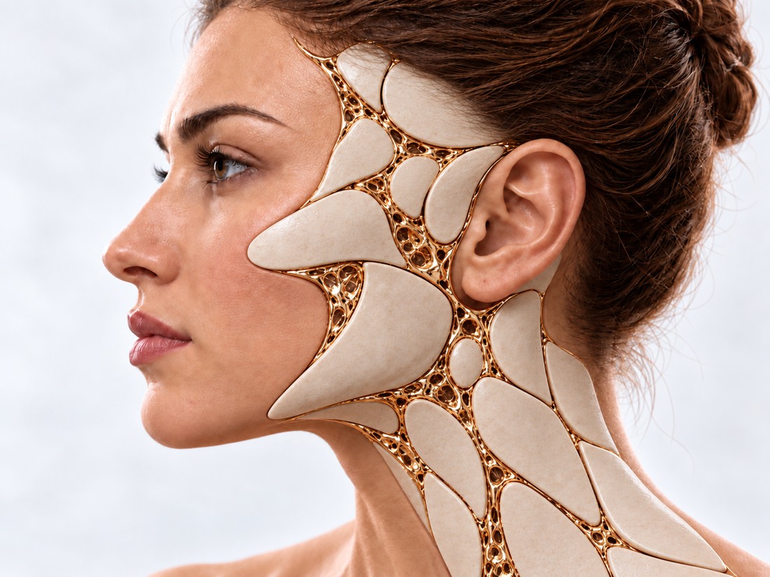
CONDITION / JAWS, MOUTH AND NECK

Mesial bite (progeny).

Mesial bite - when the lower front teeth are in front of the upper ones, because the lower jaw protrudes forward or the upper one lags behind. In children, it is corrected with orthodontic devices, in adults with a pronounced defect of the jaws - with braces along with surgery.

Conceptual visualization: jaws and biteCONCEPTUAL VISUALIZATION
What it is
The lower front teeth close in front of the upper ones; the lower jaw protrudes or the upper jaw lags behind.
How common
In a review of 53 studies, 5.93% of people with permanent teeth; this is the rarest of the three bite classes.
Who treats it
An orthodontist, and in the case of a pronounced defect of the jaw, together with a maxillofacial surgeon.
Urgency
Emergency care is not required: treatment is planned.

What is
mesial bite.

Normally, the upper front teeth slightly overlap the lower ones. With a mesial bite, the opposite is true: the lower teeth are in front, the chin protrudes, the profile becomes concave. Doctors call this a Class III bite.

The reason can be in the jaws, in the teeth, or in both: the lower jaw is too large or protrudes forward (progenia), the upper jaw is too small or located behind, or the teeth themselves are tilted. It depends on whether the braces will be enough or whether an operation is needed.

A mesial bite is a defect associated with the growth of the jaws. Therefore, in children, treatment is aimed at growth, and in adults, jaws are surgically repositioned if necessary. The opposite situation, when the upper teeth protrude above the lower ones, is a distal bite.

This is general information. The doctor will tell you what will suit you after the examination.

How
mesial bite presents.

Most of the signs are visible in the mirror and in the profile picture, and the pictures show the reason.

01

Reverse overlap

When closing, the lower front teeth stand in front of the upper ones; the position of the teeth can make chewing and brushing more difficult.

02

The chin protrudes

The lower part of the face appears more massive, the profile is concave.

03

Flat middle face

If the upper jaw lags behind, the area under the nose may appear sunken.

04

Jaw joint

Disorders of the temporomandibular joint (pain, clicking) were present in 32.5% of patients who were scheduled for jaw surgery (6 studies, 542 patients).

Dysfunction of the joint

Why is it formed?
mesial bite.

Progeny is considered the result of the interaction of genes and the environment.

Heredity

A review of 15 studies found several regions of chromosomes and genes associated with progeny, but its genetics have yet to be definitively elucidated.

Jaw growth

The lower jaw grows larger or the upper jaw smaller than it should be. Some of the changes cannot be completely corrected until the growth of the jaws is complete.

Cleft palate

After cleft surgeries, the upper jaw may lag behind. In the summary data of the literature, osteotomy of the upper jaw was required in 21% and 20.8% of patients under different treatment regimens.

Cleft palate

Teeth, not jaws

Sometimes the jaws are normal, but the teeth themselves are tilted or the lower jaw usually shifts forward when closing. It is distinguished from a real defect of the jaws on examination and photographs.

When to seek medical advice?

A mesial bite does not require emergency care - it is treated as planned. It is necessary to act urgently after surgery on the jaws.

103 immediately after jaw surgery, if:

  • difficulty breathing, speaking or swallowing;
  • swelling in the mouth or neck grows rapidly;
  • bleeding from the wound or from the mouth does not stop.

Other situations:

Immediately - to the surgeon who operated, and if it does not work - to the waiting room: pain after the operation does not weaken, but increases. On the same day, to the surgeon: a temperature of 38 °C and above or pus is released from the wound. Planned: you or your child's lower teeth close in front of the upper ones - to the orthodontist. If your teeth hurt for more than a few days after braces are installed, your gums are very red and swollen, or your braces have broken, see your orthodontist.

As defined,
what exactly to fix.

  1. Review and conversation

    How do the teeth close, does the jaw shift when closing, is there such a defect in the family, does the joint bother you.

  2. Photo and scan of teeth

    Full-face and profile photo of the face, prints or digital scan of teeth.

  3. Pictures and CT

    They show what exactly is displaced - jaws, teeth or both, and to what extent.

  4. Joint plan

    The orthodontist and the surgeon decide whether braces will be enough, whether an operation is needed and on which jaw.

How to fix
mesial bite.

The choice depends on the age, cause and severity of the defect. In children, they try to influence the growth of the jaws, in adults with a jaw defect, the bite is corrected with braces along with surgery.

01

Children

Orthodontic devices while the jaws are growing.

Face mask and other devices
The mask rests on the forehead and chin and pulls the upper jaw forward with rubber pulls. In a Cochrane review (29 studies, 1169 children aged 5–13 years), such devices significantly improved the bite immediately after treatment; the certainty of the evidence is moderate.
Is the result saved?
Long-term outcome was studied in only one study: after 3 years there was less improvement, and after 6 years - no improvement. However, doctors less often predicted the need for surgery in adulthood for children after the mask; the certainty of these data is low.
02

Adults

Braces, and for defects of the jaw - together with the operation.

Orthodontic "masking"
The orthodontist tilts the teeth so that they close without changing the jaws: the upper front ones - forward, the lower ones - back. This is an option for borderline cases, and the choice between it and surgery is not easy for them.
Orthognathic surgery
The jaws are dissected and rearranged. In a review of 6 studies with a borderline mesial bite, the operation pushed the upper jaw forward and pushed the lower jaw back — that is, it changed the jaw ratio itself.
One or two jaws
The upper jaw is pushed forward (Le Fort I osteotomy), the lower jaw is pushed back (sagittal osteotomy) or both are operated on. In a review of 9 studies, stability after 5 years or more was not significantly different; in a shorter time after the operation only on the lower jaw, the overlap of the teeth turned back a little more (a difference of 0.4 mm).
Braces before or after
Classically, first the orthodontist prepares the teeth, then the operation, then the braces stage. An operation-first approach in a review of 10 systematic reviews resulted in shorter treatment (moderate quality of evidence) but slightly more complications (low to moderate quality).
03

Risks

Numbness of the lips and chin
After surgery on the lower jaw, sensitivity may be impaired. In a review of 7 studies, according to objective tests, 63.3% on the 7th day and 12.8% after a year.
Breathing in sleep
When the lower jaw is pushed back, the airway can become narrow. In a review of 14 studies, sleep apnea after retraction of the lower jaw occurred in 8.57%, after a combination with protrusion of the upper jaw — in 0.7%, with elevation of the upper jaw — in 19.2%, mainly with greater retraction. The authors did not find any clear evidence that the reason was the operation itself.
Jaw joint
After treatment, the incidence of joint disorders in the same 6 studies did not differ from people without malocclusion; why so - it is not clear.
04

Planning and result

Virtual planning
The operation is simulated in advance on a computer based on a CT scan. In a review of 12 studies, the mean deviation from the plan was less than 2 mm in both jaws, and virtual planning appeared to be more accurate than classical planning, especially with regard to symmetry.
Individual plates
3D-planned titanium plates produced acceptable accuracy in 7 studies (74 patients); due to different evaluation methods, the authors could not give practical recommendations.
Quality of life
Dental and oral quality of life was significantly improved in both mesial and distal occlusion 4–7 months after surgery (meta-analysis of 4 studies).

How
we treat it.

A mesial bite is treated by an orthodontist and a maxillofacial surgeon together. At Dr. Valikhnovskyi's Institute of Surgery, in the "New Face" direction, the surgeon examines the bite, orders a CT scan of the facial skull and, if necessary, builds a 3D model of the jaws to assess which jaw is displaced and whether surgery is needed. Braces and braces for children are installed and managed by an orthodontist.

CLINIC SERVICE A new face Form, symmetry, facial expressions and functions after injuries, wounds, burns, removal of tumors and congenital changes. View service

What we do for this condition

  • Consultation of a maxillofacial surgeon — examination of the bite, profile and joint, talking about what is bothering you.
  • CT of the facial skull — three-dimensional images show which jaw is displaced and to what extent.
  • Virtual Planning (VSP) — a three-dimensional model of the jaws is built using a CT scan and the computer assesses where and how much they should be moved.

Rostislav Valikhnovskyi

Plastic surgeon, candidate of medical sciences, founder of Dr. Valikhnovskyi Institute of Surgery.

How treatment works with us

  1. Photos and pictures onlineSubmit full-face and profile photos, bite photos, CT scans or scans if available.
  2. ConsultationBite and joint examination, CT scan of the facial skull.
  3. 3D modelA model of the jaws is built based on the CT scan and it is shown what and how much needs to be changed.
  4. Plan with an orthodontistTogether with an orthodontist: can you do with braces or do you need surgical treatment, where to start and how many stages there will be.

Common
questions

Short answers. What is right for you, the doctor will tell you during the consultation.

Can a mesial bite be corrected without surgery?

In children, the bite is improved with orthodontic appliances, although the stability of the result has not been studied much. In adults, braces can mask the borderline shape by tilting the teeth, and the position of the jaws itself is changed by the operation.

From what age can you operate?

Decisions are made taking into account growth: some changes cannot be finally corrected until the growth of the jaws is complete. When exactly - the orthodontist and surgeon will tell after the pictures.

Is it necessary to wear braces before surgery?

The classic scheme is braces before and after surgery. There is an "surgery first" approach: in studies, treatment was shorter, but the quality of these data is low to moderate, and complications are slightly higher.

Will the lip be numb after surgery?

This is possible: sensitivity of the lower lip and chin after surgery on the lower jaw on the 7th day is impaired in 63.3%, and after a year, according to objective tests, in 12.8% (review of 7 studies).

Can surgery cause snoring or apnea?

When the lower jaw is pushed back, the airway can become narrow. Apnea after surgery was more often seen with greater displacement, in overweight and older people; the authors of the review advise overweight people to lose weight before surgery.

Are they operating on one jaw or two?

Depends on which jaw is displaced. In terms of stability after 5 years or more, no significant difference was found between surgery on one and two jaws in the studies.

Will the face change?

Yes: when the jaws are rearranged, the profile also changes. How exactly, is discussed at the consultation according to the 3D plan.

Sources

17 sources · reviews, guidelines and research
showCollapse
  1. Global distribution of malocclusion traits: A systematic reviewDental Press Journal of Orthodontics, 2018doi:10.1590/2177-6709.23.6.40.e1-10.onl ↗
  2. Orthodontic treatment for prominent lower front teeth (Class III malocclusion) in childrenCochrane Database of Systematic Reviews, 2024doi:10.1002/14651858.CD003451.pub3 ↗
  3. Developing Class III malocclusions: challenges and solutionsClinical, Cosmetic and Investigational Dentistry, 2018doi:10.2147/CCIDE.S134303 ↗
  4. Genetic Etiology in Nonsyndromic Mandibular PrognathismJournal of Craniofacial Surgery, 2017doi:10.1097/SCS.0000000000003287 ↗
  5. Orthodontic camouflage versus orthodontic-orthognathic surgical treatment in borderline class III malocclusion: a systematic reviewClinical Oral Investigations, 2022doi:10.1007/s00784-022-04685-6 ↗
  6. Stability of single-jaw vs two-jaw surgery following the correction of skeletal class III malocclusion: A systematic review and meta-analysisOrthodontics & Craniofacial Research, 2021doi:10.1111/ocr.12456 ↗
  7. Surgery-first orthognathic approach vs conventional orthognathic approach: A systematic review of systematic reviewsJournal of Stomatology, Oral and Maxillofacial Surgery, 2021doi:10.1016/j.jormas.2020.08.008 ↗
  8. Neurosensory disturbance of the inferior alveolar nerve after bilateral sagittal split osteotomy: a systematic reviewJournal of Oral and Maxillofacial Surgery, 2007doi:10.1016/j.joms.2007.05.009 ↗
  9. Influencing factors for the development of obstructive sleep apnea after orthognathic surgery in skeletal class III patients: A systematic reviewJournal of Cranio-Maxillofacial Surgery, 2024doi:10.1016/j.jcms.2024.02.014 ↗
  10. Oral Health-Related Quality of Life Changes in Patients with Dentofacial Deformities Class II and III after Orthognathic Surgery: A Systematic Review and Meta-AnalysisInternational Journal of Environmental Research and Public Health, 2022doi:10.3390/ijerph19041940 ↗
  11. Accuracy of virtual planning in orthognathic surgery: a systematic reviewHead & Face Medicine, 2020doi:10.1186/s13005-020-00250-2 ↗
  12. Accuracy of orthognathic surgery with customized titanium plates — Systematic reviewJournal of Stomatology, Oral and Maxillofacial Surgery, 2021doi:10.1016/j.jormas.2020.06.011 ↗
  13. The need for maxillary osteotomy after primary cleft surgery: A systematic review framing a retrospective studyJournal of Cranio-Maxillofacial Surgery, 2020doi:10.1016/j.jcms.2020.07.005 ↗
  14. Do patients with malocclusion have a higher prevalence of temporomandibular disorders than controls both before and after orthognathic surgery? A systematic review and meta-analysisJournal of Cranio-Maxillofacial Surgery, 2017doi:10.1016/j.jcms.2017.07.015 ↗
  15. Dental abscessNHS, 2026nhs.uk/conditions/dental-abscess ↗
  16. Cuts and grazesNHS, 2026nhs.uk/conditions/cuts-and-grazes ↗
  17. BracesNHS, 2026nhs.uk/tests-and-treatments/braces ↗

The figures on the page are from these sources.

VALIKHNOVSKI SURGERY PARTNERS

Lower jaw
comes forward?

Submit a photo of your face in your profile and a photo of your bite. The doctor will tell you whether an operation is necessary and where to start.

Call Send an enquiry
ONLINE ENQUIRY

Tell us how we can help.

Leave your contact details and briefly describe the situation. A coordinator will contact you to agree on the next step.

Preferred contact method