CONDITION / EARS

Ear microtia.

Microtia is a congenital underdevelopment of the auricle: from a slightly smaller ear of a changed shape to its complete absence. The shape of the ear is restored by surgery in childhood, and hearing is checked and treated separately - as early as possible.

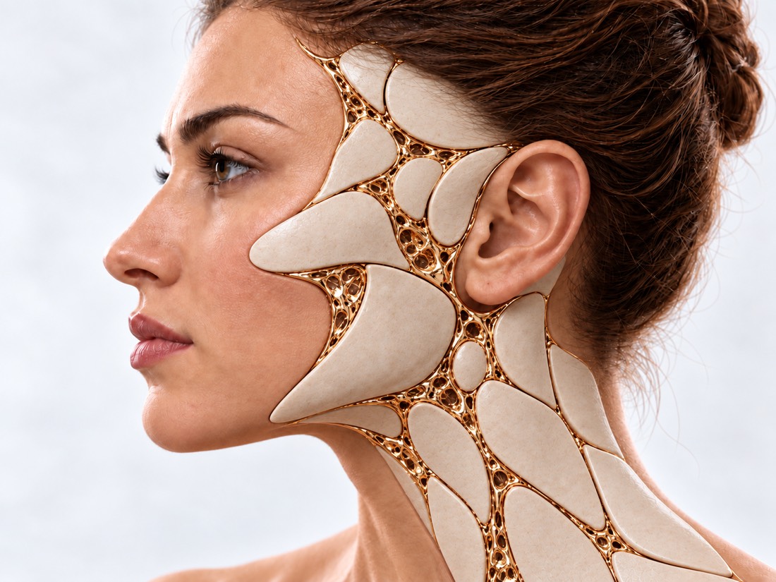
Conceptual visualization: restoration of the auricleCONCEPTUAL VISUALIZATION
What it is
The auricle is underdeveloped from birth; when it is not there at all, it is anotia.
How common
From 0.83 to 17.4 per 10,000 newborns — depending on the region.
hearing
In a study of 204 patients, the auditory canal was closed in 76% - hearing is checked as early as possible.
When surgery is performed
With an implant - usually from 3-5 years old, with own costal cartilage - later, when there is enough cartilage.

What is
microtia

Microtia is a congenital defect in which the auricle is underdeveloped. It happens in different ways: the ear is slightly smaller and without separate curls, only a ridge of skin with a lobe remains, or the ear is absent at all - this is the most severe form, anotia.

One ear is most often affected: in a study of 204 patients from the Netherlands — in 91.7%, mainly the right ear. Microtia can be a separate defect or part of a wider condition; according to a review in the Journal of Perinatology, other facial or organ malformations occur in about half of children.

The treatment has two separate parts: the hearing and the shape of the ear. Hearing is handled by an ENT doctor and a hearing specialist, and a plastic surgeon restores the shape. International consensus recommends that children with microtia and closed auditory canal be managed by a multidisciplinary team.

According to a review of studies, the moment of diagnosis is already stressful for many families. The treatment is stretched over years: the hearing is dealt with immediately, and you can calmly think about the operation on the ear.

This is general information. What will suit your child, the doctor will tell after the examination.

How it
presents.

Microtia is visible from birth. The doctor evaluates not only the auricle, but also the auditory canal, the face, and the second ear.

01

A small or altered ear

Mild degree: the auricle is smaller, part of the curls are not formed.

02

A roller with a scoop

Instead of a shell, there is a vertical ridge of skin with cartilage and a lobe. In a study of 204 patients, this type was present in 59.9% of patients with unilateral microtia.

03

Annotia

There is no auricle and lobe at all.

04

Closed auditory canal

Atresia - the auditory canal has not formed, so the child hears worse with this ear. In the same study, 76% of patients.

06

Asymmetry of the face

Underdevelopment of one half of the face — hemifacial microsomia — was present in 27.5% of patients.

Hemifacial microsomia

Why is that?
develops.

Why microtia develops and why its frequency varies so much between countries is not fully understood. Both genetic and external factors are important.

Embryo development

The ear is formed before birth. Scientists assume a violation of the development of the cells from which facial tissues are formed, or a violation of their blood supply.

Heredity

Microtia in the family: in the study of 204 patients — in 2.0%, according to the literature — in 10.0%.

Origin

Microtia is thought to be more common in Hispanics, Asians, Native Americans, and Andean people.

Concomitant defects

Facial nerve paralysis — in 8.3%, congenital heart defects — in 2.5% of patients of the same study; therefore, the child is examined completely.

Paralysis of the facial nerve

When to seek urgent medical care?

Microtia is a congenital condition, it does not require emergency care. They are urgently referred due to complications after surgery or sudden hearing loss. The main thing in the first months is to check your hearing.

103 immediately if after surgery:

  • it is difficult to breathe: the person gasps for air, cannot utter a word, the child croaks or pulls in the stomach under the ribs;
  • the chest squeezes or presses;
  • lips or skin have become very pale, blue or gray;
  • bleeding from the wound does not stop or the blood is gushing.

Other levels of urgency:

Immediately - to the surgeon who operated; if it is not possible to contact - to the reception department: after surgery, pain or swelling increases rapidly. On the same day: after the operation, the skin over the new ear or the wound on the chest is reddened, swollen, pus flows from it, the temperature rises; the skin over the frame has darkened or the frame is showing through it. On the same day - and if the child's hearing suddenly became worse or discharge appeared from the ear. Planned, in the first months: a hearing test by an ENT doctor and an audiologist, if it has not already been done - even when the other ear is healthy. Planned: consultation about the shape of the ear - long before the age of the operation, in order to calmly choose the method.

How we assess
a child

  1. Hearing test

    As early as possible, in the first weeks of life. Conventional screening of newborns does not detect all types of hearing loss, so hearing is evaluated by an audiologist in the case of microtia.

  2. Review of the ENT doctor

    Is there an auditory canal, how does the other ear hear, or is a hearing aid needed?

  3. The review is complete

    Face, jaw, movements of facial muscles, heart - because other defects occur in about half of the children.

  4. Preoperative assessment

    A photo of both ears, the size of a healthy ear and the age of the child: the method and terms depend on them.

How we treat
hearing and form.

The new ear restores its shape, but not the hearing: the hearing is treated separately and earlier. The shape is restored either with one's own costal cartilage, or with an implant, or with a prosthesis - or not at all, if the family so decides.

01

hearing

The earliest — in the first months and years.

Bone conduction apparatus
Transmits sound through the bones of the skull bypassing the closed auditory canal; babies wear it on a soft bandage. In a study of 42 children under 2 years of age, hearing with the device significantly improved and came close to normal.
Implanted device or ear canal surgery
The decision is made by the ENT doctor. In a meta-analysis of 89 studies (2,611 patients), in the immediate post-treatment period, implanted bone conduction devices improved hearing by an average of 34.4 dB, and ear canal surgery by 22.4 dB; complications were in 16.9% and 45.7%, respectively.
Why not delay
Reduced hearing can affect the development of speech and learning at school, so the selection of the device does not wait for surgery on the ear.
02

Own costal cartilage

Usually at school age.

How do they do it?
A frame is cut out of the cartilage of the ribs in the shape of a healthy ear and placed under the skin. In a study of 321 patients, 270 of whom had ear reconstruction, all who had a full ear reconstruction required at least two surgeries.
when
Many surgeons wait until 10 years: then the cartilage is enough for the frame, and the child himself agrees to the operation and helps with care.
What is known about the result
According to a review of the studies, a framework of the patient's own cartilage grew in the same way as a healthy ear, and the skin on the front surface of the ear returned to sensitivity. Some remote complications occurred in less than 10% of cases.
Breasts
After the cartilage is removed, a scar remains and the chest can change shape: in studies - 0% to 50%, and these figures are probably underestimated. Preservation of the cartilage lining (cartilage) reduced the risk. Among other complications in the area of ​​the intake are a hematoma, damage to the pleura (lung lining) and a keloid scar; in a study of 321 patients, they were few.
03

Implant

Earlier, usually from 3-5 years.

Porous polyethylene
The finished frame does not require the removal of ribs, so the operation can be done at a younger age.
Risks
In a meta-analysis of 11 studies (3,816 patients), with an implant, the framework was more often opened — a difference of 6.97 percentage points; the authors write that the implant may also be associated with more frequent infection and repeat operations, but the quality of the studies is low.
satisfaction
In another meta-analysis (68 studies, 5,546 children), post-cartilage and post-implant satisfaction was similar at over 90%.
04

Without reconstruction

Follow-up
Surgery is optional: some families choose only hearing treatment.
Prosthesis on bone implants
Silicone ear prosthesis is attached to titanium pins in the skull bone. In a study of 27 patients (more than half after tumor treatment) with prostheses of the ear, nose and eye area, the success rate was 86%, and 2 patients (7.4%) could not use the prosthesis.
Child support
For the most part, children with microtia and microsomia develop within normal limits, but difficulties with communication and speech are possible; A review of research suggests psychological support and help with learning.

How
we treat it.

At Dr. Valikhnovskyi's Institute of Surgery, the shape of the ear in case of microtia and anotia is restored in the direction of "Growing the ear". Hearing is checked and treated by an ENT doctor and an audiologist - the clinic is responsible for the reconstruction of the auricle and coordinates it with hearing treatment.

CLINIC SERVICE We grow an ear Auricle with microtia and anotia, after injuries, burns and removal of tumors, consequences of unsuccessful otoplasty. View service

What we do for this condition

  • Reconstruction with own costal cartilage — the frame is cut from the cartilage of the ribs according to the pattern of a healthy ear; in 2 stages or more.
  • Reconstruction with a Medpor implant — the frame made of porous polyethylene is wrapped with its own fascia of the temple; often in 1 stage.
  • 3D templates and mirror modeling — a healthy ear is scanned and a mirror model is printed, with which the frame is compared.
  • Epiprosthesis on titanium implants — silicone ear prosthesis with magnets or clips — when surgical reconstruction is impossible.

Rostislav Valikhnovskyi

Plastic surgeon, candidate of medical sciences, founder of Dr. Valikhnovskyi Institute of Surgery.

How treatment works with us

  1. Photo onlineSend a photo of both ears directly and from the side and the results of the hearing test - the doctor will pre-evaluate the situation.
  2. ConsultationExamination of the ear and face, discussion of methods and age of surgery; an adult child is also asked.
  3. PlanCartilage or implant, number of stages and coordination with hearing treatment.
  4. OperationsThe frame is compared to a 3D model of a healthy ear; each stage in its own time.
  5. Follow-upReviews between stages and after; according to a review of studies, observation is advised for at least 5 years.

Common
questions

Short answers. What is right for you, the doctor will tell you during the consultation.

From what age is the operation performed?

With an implant - usually from 3-5 years. Own rib cartilage - later, when the rib cartilage is sufficient: many surgeons wait up to 10 years. The doctor names the specific age after the examination.

Which is better: costal cartilage or an implant?

Studies do not give a clear answer. The cartilage grows with the baby, but leaves a scar on the chest and requires at least two surgeries. The implant is placed earlier and without taking the ribs, but the frame is opened more often. The choice is discussed individually.

Will hearing improve after reconstruction?

No, reconstruction restores the shape of the ear. An ENT doctor treats hearing separately - with a bone conduction device, an implanted device or an operation on the auditory canal.

The second ear is healthy. Do you need a device?

This is decided by an ENT doctor and a hearing specialist after a hearing test. In a study of 42 children, a bone conduction device improved hearing in both unilateral and bilateral hearing loss.

Is it necessary to operate?

No. You can only treat the hearing, choose a prosthesis or return to the question when the child grows up and can decide together with the parents.

Will the shape of the breast change after cartilage removal?

Maybe: in research - from 0% to 50%. The risk is lower when the surgeon preserves the cartilage. The scar on the chest remains.

What to do if it becomes difficult to breathe or the pain increases after the operation?

If it is difficult to breathe, the lips are blue or the bleeding does not stop, call 103 immediately. If the pain or swelling increases quickly, contact the surgeon who operated immediately, and if that does not work, go to the emergency room. Redness, pus or fever is a reason to see a surgeon the same day.

Sources

19 sources · international consensus, meta-analyses, systematic reviews, research and NHS
showCollapse
  1. International Consensus Recommendations on Microtia, Aural Atresia and Functional Ear ReconstructionThe Journal of International Advanced Otology, 2019doi:10.5152/iao.2019.7383 ↗
  2. Rib Autograft Versus Porous Polyethylene Implant Outcomes in Microtia Reconstruction: A Meta-Analysis and Systematic ReviewThe Cleft Palate Craniofacial Journal, 2026doi:10.1177/10556656251349274 ↗
  3. Reconstructive Techniques in Pediatric Congenital Microtia: A Systematic Review and Meta-analysisFacial Plastic Surgery, 2025doi:10.1055/a-2247-5109 ↗
  4. Long-term complications of microtia reconstruction: A systematic reviewJournal of Plastic, Reconstructive & Aesthetic Surgery, 2021doi:10.1016/j.bjps.2021.08.001 ↗
  5. Long-term aesthetics, patient-reported outcomes, and auricular sensitivity after microtia reconstruction: A systematic reviewJournal of Plastic, Reconstructive & Aesthetic Surgery, 2021doi:10.1016/j.bjps.2021.08.004 ↗
  6. Comprehensive analysis of donor-site chest deformities after autologous costal cartilage microtia reconstruction: A systematic reviewJournal of Plastic, Reconstructive & Aesthetic Surgery, 2024doi:10.1016/j.bjps.2024.05.005 ↗
  7. Microtia: Epidemiology and geneticsAmerican Journal of Medical Genetics Part A, 2012doi:10.1002/ajmg.a.34352 ↗
  8. Microtia in the Netherlands: Clinical characteristics and associated anomaliesInternational Journal of Pediatric Otorhinolaryngology, 2014doi:10.1016/j.ijporl.2014.03.024 ↗
  9. Microtia and Related Facial AnomaliesClinics in Perinatology, 2018doi:10.1016/j.clp.2018.07.007 ↗
  10. Comparison of Outcomes of Surgery Versus Implantable Device for the Treatment of Hearing Loss Associated With Congenital Aural Atresia: A Systematic Review and Meta-AnalysisOtology & Neurotology, 2023doi:10.1097/MAO.0000000000003950 ↗
  11. Effectiveness of Bone Conduction Hearing Aids in Young Children with Congenital Aural Atresia and MicrotiaMedical Science Monitor, 2021doi:10.12659/MSM.933915 ↗
  12. Psychosocial and Health-Related Experiences of Individuals With Microtia and Craniofacial Microsomia and Their Families: Narrative Review Over 2 DecadesThe Cleft Palate Craniofacial Journal, 2023doi:10.1177/10556656221091699 ↗
  13. General and Treatment-Specific Outcomes with Osseointegrated Implants in Auricular, Nasal, and Orbital Prosthetic ReconstructionCraniomaxillofacial Trauma & Reconstruction, 2025doi:10.3390/cmtr18010016 ↗
  14. Autologer Rippenknorpel in der Rhino- und Ohrmuschelchirurgie: Komplikationen anhand von 312 FällenLaryngo-Rhino-Otologie, 2013doi:10.1055/s-0033-1351261 ↗
  15. Ear MicrotiaStatPearls, 2024pubmed.ncbi.nlm.nih.gov/33085390 ↗
  16. Hearing lossNHS, 2025nhs.uk/conditions/hearing-loss ↗
  17. Newborn hearing screeningNHS, 2026nhs.uk/baby/newborn-screening/hearing-test ↗
  18. Shortness of breathNHS, 2024nhs.uk/symptoms/shortness-of-breath ↗
  19. Cuts and grazesNHS, 2026nhs.uk/conditions/cuts-and-grazes ↗

The figures on the page are from these sources.

VALIKHNOVSKI SURGERY PARTNERS

The child was born
with microtia?

Send a photo of both ears and hearing test results. The doctor will tell you what the options are and when it is better to discuss them.

Call Send an enquiry
ONLINE ENQUIRY

Tell us how we can help.

Leave your contact details and briefly describe the situation. A coordinator will contact you to agree on the next step.

Preferred contact method