CONDITION / FACE

Neurofibromas of the head and face.

Neurofibroma is a benign tumor of the nerve sheath. On the face and head, multiple neurofibromas are most often associated with neurofibromatosis type I (NF1); whether they interfere or not depends on the size and location.

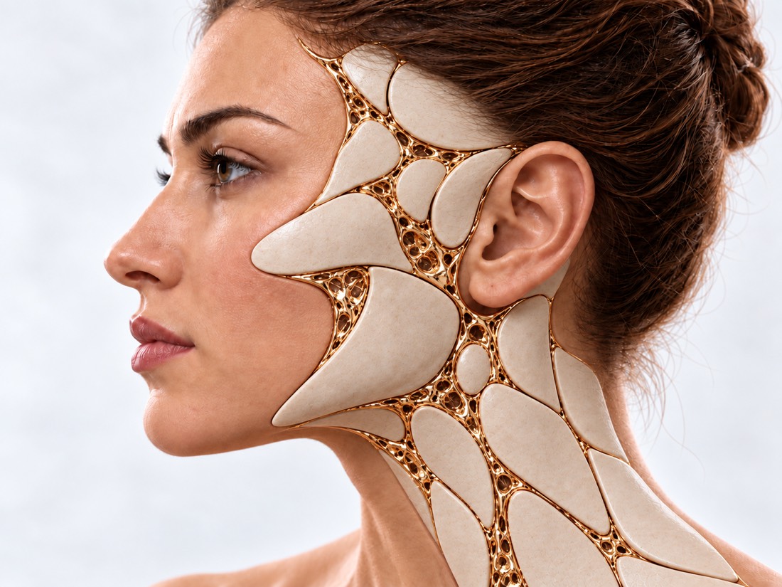
Conceptual visualization: restoration of face and head contours after removal of tumorsCONCEPTUAL VISUALIZATION
What it is
Benign nerve sheath tumors: nodules on the skin or large plexiform tumors.
How common
About 1 in 1,900 to 3,500 people have neurofibromatosis type I.
risk
According to a population-based study in England, 8–13% of people with NF1 develop a malignant nerve sheath tumor (MPNST) during their lifetime.
Who treats it
A surgeon removes tumors and restores tissues; supervision and medication are conducted by a geneticist or an oncologist.

What is
neurofibromas.

Neurofibromas are benign tumors that grow on nerves and in the skin. Multiple neurofibromas are the main feature of neurofibromatosis type I (NF1), a hereditary disease in which a person has an increased susceptibility to tumors.

Doctors distinguish between two main types. Cutaneous neurofibromas are nodules on or under the skin; they appear in almost all people with NF1, usually in their late teens or early 20s. Plexiform neurofibromas grow along nerve trunks, are large and deep; on the face, they change the contour and symmetry, can hurt or weaken the muscles.

Neurofibromas are benign, but people with NF1 can develop a malignant tumor of the nerve sheath — doctors call it MPNST. In a population-based study in England, the lifetime risk was 8–13%, and half of these tumors were found in people younger than 26 years. Therefore, neurofibromas are monitored throughout life.

NF1 is a lifelong condition that progresses differently for everyone. There is no medicine that can cure it, but the tumors that interfere can be removed, and some of the plexiform ones can be reduced with medicine.

This is general information. The doctor will tell you what will suit you after the examination.

How they manifest
neurofibromas.

Symptoms depend on where the tumor is and how big it is. Many people with NF1 have moderate symptoms.

01

Nodules on the skin

Soft nodules on or under the skin that sometimes hurt or itch. There can be several to hundreds of them.

02

A large tumor

Plexiform neurofibroma is a mild thickening of tissue along a nerve that changes the contour and symmetry of the face.

03

Pain and weakness

The tumor presses on the nerve: pain, muscle weakness, and movement disorders appear.

04

eyes

Strabismus, worse vision, or a bulging eye — when the tumor is close to the eye socket or optic nerve.

Neoplasms of the eyelids
05

Spots and freckles

Light brown spots the color of coffee with milk and freckles under the armpits are signs that suspect NF1.

06

Alarming changes

The tumor is growing rapidly, pain is increasing, new weakness or numbness has appeared - this should be shown to the doctor.

Why do they appear?
neurofibromas.

The cause of NF1 is a change in one gene. What affects the growth of tumors is partially known.

NF1 gene

The NF1 gene inhibits the growth of tumors. When one copy of it doesn't work, neurofibromas form more easily in the tissues.

Heredity

A person with NF1 will pass on the altered gene to each child with a 50% chance. But a child can be born with NF1 even if the parents do not have it.

age

Plexiform neurofibromas grow most actively in childhood. In a study of 201 patients, 13.5% of tumors significantly increased, more often in children, and 35.5% decreased over time.

pregnancy

During pregnancy, more neurofibromas may appear, or existing ones may become larger and itchy.

When to seek medical advice?

Neurofibromas usually grow slowly and do not require emergency treatment. But it is important not to miss signs that the tumor is pressing on the eye or respiratory tract, or that it could become malignant.

103 immediately if:

  • suddenly stopped seeing in one or both eyes;
  • sudden severe pain in the eye;
  • it is very difficult to breathe: a person gasps for air with his mouth, suffocates or cannot utter words.

Do not delay a visit to the doctor:

On the same day: the headache does not go away or worsens, you are constantly nauseous or vomiting, your vision has become blurred or worsened. In the coming days: neurofibroma grows rapidly, pain increases, weakness or numbness gradually appear - this is how they check whether the tumor has become malignant. Planned: new nodules, change in facial appearance, desire to remove neurofibromas. People with NF1 are advised to have an examination at least once a year, an eye test for children - every year, and for adults - every 2 years.

As defined,
what to treat

  1. Discussion and review

    When the tumors appeared, do they grow, do they hurt; does NF1 run in the family. The doctor examines the skin: spots, freckles, nodules.

  2. Genetic analysis

    Current criteria for the diagnosis of NF1 combine physical examination and genetic testing. It is prescribed by a geneticist.

  3. MRI

    Shows how deep the plexiform neurofibroma goes and what structures it affects.

  4. Ophthalmologist's examination

    Vision and eye testing is a mandatory part of NF1 follow-up.

  5. Histology

    If the tumor grows quickly or hurts, the doctor prescribes examination and examination of the tissue under a microscope to rule out malignancy. If a malignant tumor is suspected, treatment is planned together with an oncologist.

How we treat
neurofibromas.

If the neurofibromas do not interfere, the doctor may suggest observation. It is treated when the tumor hurts, disrupts function, grows quickly or noticeably changes the face.

01

Cutaneous neurofibromas

Nodules on the skin.

Removal
The main method is to surgically remove the nodules. There are currently no drugs that would act specifically on cutaneous neurofibromas.
Laser
The CO2 laser can remove many nodules at once, but often leaves scars and light or dark spots.
02

Plexiform neurofibromas

Operation.

When surgery is performed
To exclude a malignant tumor, if it increases rapidly; to reduce pain or weakness; if the tumor presses on the respiratory tract; to improve the appearance of the face.
In whole or in part
Only a small tumor can usually be completely removed. In a study of 39 children, a large tumor grew back after surgery in 18 of 18, an average of 3.1 years later.
The face is more complicated
Tumors of the head and face grow back more often than tumors of the extremities; as well as in children under 10 years old and when the tumor cannot be removed almost completely.
After surgery
In the cohort of 52 patients, early complications were in 16%, late complications in 13%; 19% with residual tumor were operated on again. 14 completely removed tumors did not return during the observation period (2.9 years on average).
03

Medicines

Selumetinib is for tumors that cannot be removed.

What it is
Oral drugs that inhibit the growth of plexiform neurofibroma cells. A geneticist or an oncologist prescribes and conducts treatment.
Children
In a study of 50 children, the tumor was significantly reduced by 70%, and in most of them the effect lasted for more than a year.
Adults
In a randomized trial, tumors shrank in 20% of adults on selumetinib versus 5% on placebo.
Side effects
Most often, nausea, vomiting or diarrhea, acne-like rash, inflammation near the nails.
04

Supervision

All life
Regular examinations help to notice new tumors, vision changes and alarming signs in time. Guidelines recommend managing people with NF1 in a specialist center with different specialists.

How
we treat it.

Neurofibromas of the head and face are treated at Dr. Valikhnovskyi's Institute of Surgery in the "New Face" direction: the clinic is responsible for removing tumors and restoring tissues. NF1 diagnosis, genetic counseling, medication, and lifelong follow-up are handled by a geneticist or oncologist—we work with them.

CLINIC SERVICE A new face Form, symmetry, facial expressions and functions after injuries, wounds, burns, removal of tumors and congenital changes. View service

What we do for this condition

  • Tumor removal with local flaps — small and medium defects cover the face and neck with tissues — they are the closest in color and texture.
  • Perforating facial flaps — a flap of skin on a thin vascular pedicle closes the wound without gross tension and distortion of the lines of the mouth or eyes.
  • Fabric dermotension (expander) — a supply of healthy skin is grown next to the tumor, and after the tumor is removed, it is moved to the defect.
  • Free microsurgical flaps — after removal of the tumor, large defects are replaced with tissues from other parts of the body, suturing the vessels under a microscope.
  • Lipofilling — own fat helps to smooth the contour of the face after major reconstructions.

Rostislav Valikhnovskyi

Plastic surgeon, candidate of medical sciences, founder of Dr. Valikhnovskyi Institute of Surgery.

Publications by topic

  • "Surgical tactics and results of treatment of neurofibrosis of the head: clinical experience" — Advances in clinical and experimental medicine, 2025 (DOI).

How treatment works with us

  1. Photo onlineSend a photo of the face directly and from the side and, if available, an MRI and a genetic report.
  2. ConsultationExamination, assessment of MRI; if necessary, referral to a geneticist or oncologist.
  3. PlanWhat to remove, completely or partially, how to close the defect and whether it is necessary to discuss medication first.
  4. SurgeryTumor removal with tissue restoration; removed must be examined under a microscope.
  5. SupervisionPost-operative examinations, followed by regular follow-up for NF1.

Common
questions

Short answers. What is right for you, the doctor will tell you during the consultation.

Is neurofibroma cancer?

No, neurofibroma is benign. But in people with NF1, the risk of a malignant tumor of the nerve sheath during life, according to a study in England, is 8-13%. Its signs are rapid tumor growth, increasing pain, new weakness or numbness.

Can plexiform neurofibroma be removed completely?

A small one can sometimes be removed completely. A large one is usually partially removed and can grow back, so the plan is based on size, location, and age.

Will new neurofibromas appear after surgery?

Skin nodules may continue to appear: NF1 is a lifelong condition. A plexiform tumor that has not been completely removed can grow back, so follow-up is necessary after surgery.

Is it possible to treat with drugs instead of surgery?

Selumetinib is prescribed when a plexiform neurofibroma cannot be safely removed. It shrinks the tumor in some patients but does not cure NF1. Medicines are administered by a geneticist or an oncologist.

Will NF1 be passed on to children?

The probability of passing on the changed gene to each child is 50%. You should talk to a geneticist about family planning.

At what age is it better to operate on a child?

Tumors grow more actively in children, and in younger children, they return more often after surgery. Therefore, the decision is made individually, considering how much the tumor interferes now.

Cutaneous neurofibromas — scalpel or laser?

Both ways are possible. The CO2 laser removes many nodules at a time, but more often leaves scars and blemishes. Surgical removal can give a good cosmetic result, but with a large number of nodules, anesthesia may be required.

Sources

18 sources · guidelines, consensus, clinical trials and NHS
showCollapse
  1. Revised diagnostic criteria for neurofibromatosis type 1 and Legius syndrome: an international consensus recommendationGenetics in Medicine, 2021doi:10.1038/s41436-021-01170-5 ↗
  2. Care of adults with neurofibromatosis type 1: a clinical practice resource of the American College of Medical Genetics and Genomics (ACMG)Genetics in Medicine, 2018doi:10.1038/gim.2018.28 ↗
  3. ERN GENTURIS tumour surveillance guidelines for individuals with neurofibromatosis type 1EClinicalMedicine, 2023doi:10.1016/j.eclinm.2022.101818 ↗
  4. Neurofibromatosis 1 French national guidelines based on an extensive literature review since 1966Orphanet Journal of Rare Diseases, 2020doi:10.1186/s13023-020-1310-3 ↗
  5. Management of neurofibromatosis type 1-associated plexiform neurofibromasNeuro-Oncology, 2022doi:10.1093/neuonc/noac146 ↗
  6. Selumetinib in Children with Inoperable Plexiform NeurofibromasNew England Journal of Medicine, 2020doi:10.1056/NEJMoa1912735 ↗
  7. Efficacy and safety of selumetinib in adults with neurofibromatosis type 1 and symptomatic, inoperable plexiform neurofibromas (KOMET): a multicentre, international, randomised, placebo-controlled, parallel, double-blind, phase 3 studyLancet, 2025doi:10.1016/S0140-6736(25)00986-9 ↗
  8. Malignant peripheral nerve sheath tumours in neurofibromatosis 1Journal of Medical Genetics, 2002doi:10.1136/jmg.39.5.311 ↗
  9. Management of head and neck plexiform neurofibromas in pediatric patients with neurofibromatosis type 1Archives of Otolaryngology–Head & Neck Surgery, 2005doi:10.1001/archotol.131.8.712 ↗
  10. Prognostic signs in the surgical management of plexiform neurofibroma: the Children's Hospital of Philadelphia experience, 1974-1994Journal of Pediatrics, 1997doi:10.1016/s0022-3476(97)70092-1 ↗
  11. Growth behavior of plexiform neurofibromas after surgeryGenetics in Medicine, 2013doi:10.1038/gim.2013.30 ↗
  12. Growth dynamics of plexiform neurofibromas: a retrospective cohort study of 201 patients with neurofibromatosis 1Orphanet Journal of Rare Diseases, 2012doi:10.1186/1750-1172-7-75 ↗
  13. Management of cutaneous neurofibroma: current therapy and future directionsNeuro-Oncology Advances, 2020doi:10.1093/noajnl/vdz034 ↗
  14. Evaluation and Factors of Quality of Life Among Patients With Neurofibromatosis Type 1-Associated Craniofacial Plexiform NeurofibromasJournal of Craniofacial Surgery, 2020doi:10.1097/SCS.0000000000006015 ↗
  15. Neurofibromatosis type 1 (NF1)NHS, 2025nhs.uk/conditions/neurofibromatosis-type-1 ↗
  16. Vision lossNHS, 2025nhs.uk/conditions/vision-loss ↗
  17. Shortness of breathNHS, 2024nhs.uk/symptoms/shortness-of-breath ↗
  18. Surgical tactics and results of treatment of neurofibromas of the head: clinical experienceAchievements of clinical and experimental medicine, 2025doi:10.11603/1811-2471.2025.v.i3.15555 ↗

The figures on the page are from these sources.

VALIKHNOVSKI SURGERY PARTNERS

Neurofibromas grow
do they change faces?

Send a photo of your face and MRI if available. The doctor will tell you what can be removed and how to restore the tissues.

Call Send an enquiry
ONLINE ENQUIRY

Tell us how we can help.

Leave your contact details and briefly describe the situation. A coordinator will contact you to agree on the next step.

Preferred contact method