CONDITION / FACE

Nevi (moles) on the face and head.

Almost everyone has facial moles, or nevi, and most of them are harmless. Another important thing is to notice the changes that will make the doctor suspect melanoma, and to remove the birthmark so that it can be examined.

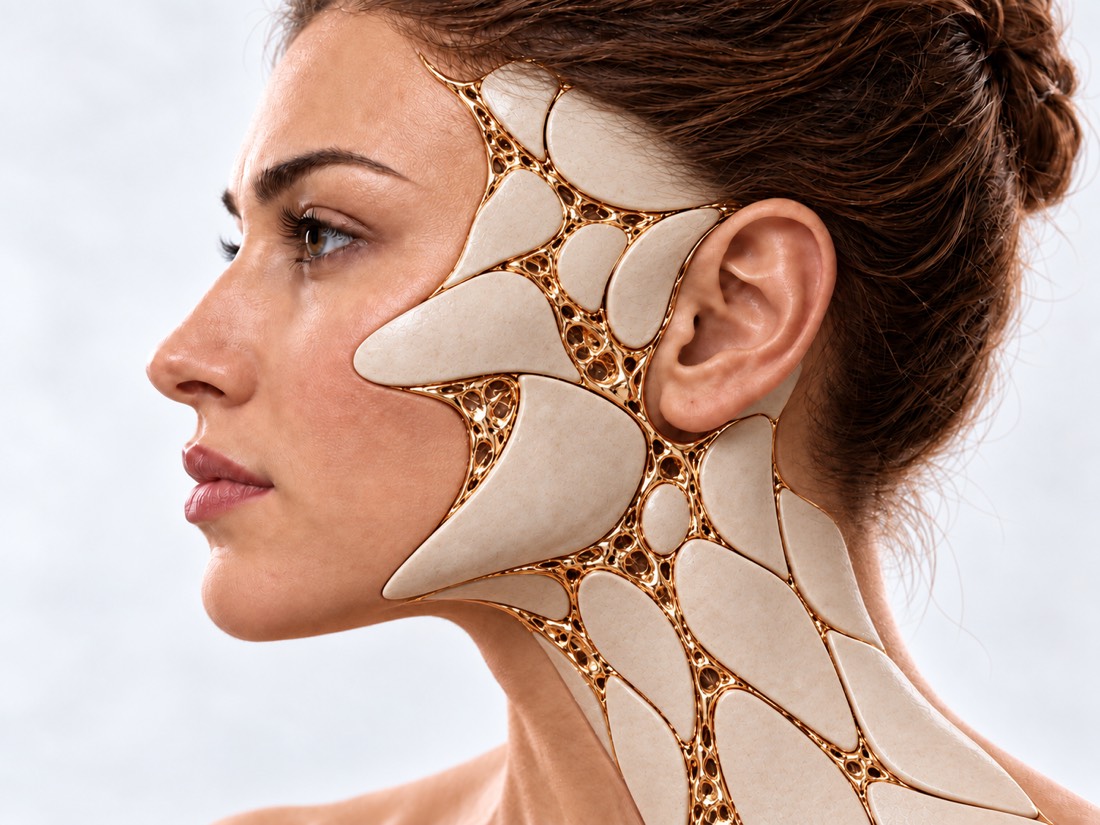
Conceptual visualization: skin of the face and headCONCEPTUAL VISUALIZATION
What it is
A benign accumulation of pigmented skin cells is a spot or bump.
What worries
A change in size, shape, or color may be a sign of melanoma.
It is important to know
Only 29.1% of melanomas arise from an existing mole, the rest - on intact skin.
Who treats it
Evaluated by a dermatologist, removed by a surgeon; melanoma is treated by an oncologist.

What is
nevus

A nevus, or birthmark, is a cluster of pigmented skin cells. Moles are flat or convex, smooth or rough, sometimes hair grows from them. They are present in almost everyone and mostly do not require treatment.

Some birthmarks are congenital: they are already in the baby. Others appear throughout life, especially during childhood and adolescence. It is normal if moles fade or disappear with age, and become slightly darker during pregnancy.

The main question about any mole is whether it is melanoma. This is the most dangerous skin tumor: it accounts for 90% of skin cancer deaths. At the same time, most melanomas do not arise from a mole: in a meta-analysis of 38 studies, only 29.1% of melanomas were based on an existing nevus, and 70.9% appeared on intact skin. Therefore, they monitor both old birthmarks and new spots.

A special type of melanoma occurs on the face of older people - malignant lentigo. It is a slowly growing light or dark brown spot with uneven edges on skin that has been damaged by the sun for years.

This is general information. The doctor will tell you what will suit you after the examination.

How to notice
a disturbing birthmark.

Doctors use the ABCDE rule — these are the five signs by which a mole should be shown to a doctor. Another clue is a mole that is clearly not like the rest of yours.

01

A is asymmetry

The halves of the mole are different in shape. Normal birthmarks are mostly round or oval.

02

B - edges

The edges are uneven, "torn" or blurred. A normal birthmark has a flat edge.

03

C is color

Two colors and more. A normal birthmark is usually one color.

04

D is the diameter

More than 6 mm. But early melanoma can also be smaller, so all signs weigh together.

05

E - changes

The birthmark grows, changes shape or color, itches, hurts, bleeds or crusts over. Doctors recommend paying special attention to changes.

Melanoma of the skin of the face
06

"The Ugly Duckling"

A birthmark that is clearly different from others on this person. In the study, nine dermatologists labeled all melanomas in the sample as such.

Where do they come from?
moles

Moles appear without any reason. But the risk of melanoma depends on their number, type and sun exposure.

Congenital nevi

Is from birth. In a review of 7,915 patients, melanoma developed in 1.15%; in large and giant nevi, the risk was 21.9 times higher than in small and medium ones.

Giant congenital nevus

Acquired birthmarks

They appear throughout life, mostly in childhood and adolescence. New birthmarks at this age are the norm.

Many moles

The more of them, the higher the risk of melanoma: 101-120 moles versus less than 15 increased it 6.89 times, and 5 atypical moles versus none increased it 6.36 times.

the sun

The sun's ultraviolet rays increase the likelihood that a mole will become malignant. Solarium also gives ultraviolet.

When to seek medical advice?

Moles by themselves do not require emergency care. But changes in the birthmark are a reason not to postpone the examination: melanoma that was found early is easier to treat.

103 immediately if:

  • the birthmark was injured, and the bleeding does not stop, although you press the wound tightly;
  • the blood spurts, bright red and hard to stop.

See a doctor without delay if:

On the same day: the injured birthmark is reddened, swollen, hurts more and more, or pus is released from it. In the coming days: the mole has changed in size, shape or color, itches, hurts, is inflamed, bleeds or crusted over; a new unusual spot has appeared that does not go away for several weeks. If the doctor suspects melanoma, you should see an oncologist or dermato-oncologist within 2 weeks. Planned: the birthmark doesn't change, but it bothers you or you don't like the way it looks.

How to evaluate
birthmark

  1. Conversation

    When did the birthmark appear and did it change; have you or a family member had melanoma or other skin cancer.

  2. Skin examination

    The doctor examines the mole and the rest of your skin and compares it to your other moles.

  3. Dermatoscopy

    View through a magnifying device with illumination. In a Cochrane review, it recognized 92% of melanomas with conventional screening versus 76% without it.

  4. Follow-up

    People at high risk have repeat digital dermatoscopies and full-body photos taken to detect changes early.

  5. Histology

    The final answer is given by examination of the tissue under a microscope. If melanoma is suspected, it is always needed.

How to delete
moles

It is not necessary to treat an ordinary birthmark. It is removed if it causes suspicion or interferes with the view. The main rule is to examine the removed tissue under a microscope.

01

When to delete

Decisions are made after examination and dermatoscopy.

Melanoma is suspected
The mole is removed completely and must be examined: this is the only way to make a diagnosis.
In appearance
Benign moles on the face are often removed at the request of a person, because they interfere with the appearance.
Congenital nevus
The decision depends on size, location and age. Large nevi are removed in stages, often with an expander.
02

Methods

The scar and whether the mole will return depend on the method.

Excision with suture
The birthmark is cut out together with a thin strip of healthy skin and sewn up. In a randomized trial, moles did not return after excision, but the scar was larger than after excision.
Cutting
The convex mole is cut at the level of the skin without stitches. The scar is smaller, but nevus cells remain more often: in the same study, the mole returned in 11.7%, in another series of 204 moles - in 19.6%.
Laser
In a review of studies of congenital nevi, the laser briefly lightened the pigment, but often left a scar and the pigment returned. There are no reliable data on long-term safety.
03

If it is melanoma

Oncologist
Treatment is conducted by an oncologist or a dermato-oncologist: based on histology, he determines the stage, examination and limits of re-excision.
Examination
Whether additional examinations are required depends on the thickness of the melanoma and the histological stage; they are prescribed by an oncologist.
Lentigo on the face
This type of melanoma can spread beyond visible boundaries, so the boundaries of removal are planned especially carefully, sometimes in stages.
Supervision
After treatment, regular check-ups are needed to detect recurrence or new melanoma early.

How
we treat it.

Moles on the face and head at Dr. Valikhnovskyi's Institute of Surgery are removed in the "New Face" direction - so that the tissue can be examined, and the wound can be closed taking into account the facial features. If histology shows melanoma, its treatment is led by an oncologist; the clinic is responsible for tissue removal and recovery.

CLINIC SERVICE A new face Form, symmetry, facial expressions and functions after injuries, wounds, burns, removal of tumors and congenital changes. View service

What we do for this condition

  • Removal with histology — skin neoplasms are removed and examined; what to do next depends on the conclusion.
  • Local patches - if a larger defect remains after removal, it is covered with skin from the face or neck - it is the closest in color and texture.
  • Perforating facial flaps — allow you to close the wound without rough tension and without distorting the lines of the mouth or eyes.
  • Expander — in the case of a large nevus, the nearby healthy skin is stretched and moved to the place of the removed one.
  • Reconstruction in one operation — if the cut edges are clean, the defect is closed immediately; if not, after histology.

Rostislav Valikhnovskyi

Plastic surgeon, candidate of medical sciences, founder of Dr. Valikhnovskyi Institute of Surgery.

How treatment works with us

  1. Photo onlineSend clear photos of the mole from up close and from afar - the doctor will assess the situation in advance.
  2. ConsultationExamination of mole and skin; if necessary, consult a dermatologist.
  3. PlanIs it necessary to remove, in what way and how to close the wound.
  4. RemovalThe birthmark is removed and sent for histological examination.
  5. Conclusion and recoveryThe doctor explains the result of the histology and monitors healing; for melanoma refers to an oncologist.

Common
questions

Short answers. What is right for you, the doctor will tell you during the consultation.

Is it possible to remove a birthmark that does not change?

It is possible, if it interferes. Removal becomes mandatory when there are signs according to ABCDE: then the tissue is examined under a microscope.

Can every birthmark become melanoma?

Most melanomas appear on intact skin: in a meta-analysis of 38 studies, 29.1% arose from a mole. Therefore, it is important to monitor both old moles and new spots.

Why research a removed mole?

Only under a microscope can you say for sure that it is benign. In a study of 33,280 normal skin samples, melanomas also occurred — 0.16%, some of which appeared to be benign.

Can a mole be removed with a laser?

For questionable birthmarks, removal with histology is chosen. With congenital nevi, the laser often left a scar, and the pigment returned; there are no reliable data on long-term safety.

Will there be a scar?

Yes, its size depends on the method of removal. In a series of 204 excised birthmarks, the imperceptible scar was more often left on the face.

A birthmark is stuck and bleeding - what to do?

Press firmly with a clean tissue or bandage. If the bleeding doesn't stop, call 103. Then show the mole to a doctor — especially if it started bleeding on its own.

How to protect moles on the face?

Hat and sunglasses, cream with SPF at least 30, no tanning beds. And regularly examine the skin to notice changes in time.

Sources

17 sources · reviews, guidelines and research
showCollapse
  1. European consensus-based interdisciplinary guideline for melanoma. Part 1: Diagnostics - Update 2024European Journal of Cancer, 2025doi:10.1016/j.ejca.2024.115152 ↗
  2. Guidelines of care for the management of primary cutaneous melanomaJournal of the American Academy of Dermatology, 2019doi:10.1016/j.jaad.2018.08.055 ↗
  3. Early diagnosis of cutaneous melanoma: revisiting the ABCD criteriaJAMA, 2004doi:10.1001/jama.292.22.2771 ↗
  4. Ugly Duckling Sign as a Major Factor of Efficiency in Melanoma DetectionJAMA Dermatology, 2017doi:10.1001/jamadermatol.2016.5500 ↗
  5. Dermoscopy, with and without visual inspection, for diagnosing melanoma in adultsCochrane Database of Systematic Reviews, 2018doi:10.1002/14651858.CD011902.pub2 ↗
  6. A meta-analysis of nevus-associated melanoma: Prevalence and practical implicationsJournal of the American Academy of Dermatology, 2017doi:10.1016/j.jaad.2017.06.149 ↗
  7. Meta-analysis of risk factors for cutaneous melanoma: I. Common and atypical naeviEuropean Journal of Cancer, 2005doi:10.1016/j.ejca.2004.10.015 ↗
  8. Risk of melanoma in congenital melanocytic nevi of all sizes: A systematic reviewJournal of the European Academy of Dermatology and Venereology, 2023doi:10.1111/jdv.18581 ↗
  9. Surgery or Not? A Systematic Review of Facial Congenital Melanocytic Nevi Treatment Patterns and OutcomesAnnals of Plastic Surgery, 2024doi:10.1097/SAP.0000000000003676 ↗
  10. Laser treatment of congenital melanocytic naevi: a systematic reviewBritish Journal of Dermatology, 2018doi:10.1111/bjd.16094 ↗
  11. Shave excision of common acquired melanocytic nevi: cosmetic outcome, recurrences, and complicationsDermatologic Surgery, 2005doi:10.1097/00042728-200509000-00005 ↗
  12. Shave Excision Versus Elliptical Excision of Nonpigmented Intradermal Melanocytic Nevi: Comparative Assessment of Recurrence and Cosmetic OutcomesDermatologic Surgery, 2021doi:10.1097/DSS.0000000000002666 ↗
  13. Unexpected high-risk neoplasias among routine skin specimensDanish Medical Journal, 2026doi:10.61409/A09250776 ↗
  14. Lentigo maligna part I: Epidemiology, risk factors, and diagnosisJournal of the American Academy of Dermatology, 2025doi:10.1016/j.jaad.2024.09.065 ↗
  15. MolesNHS, 2023nhs.uk/conditions/moles ↗
  16. Symptoms of melanoma skin cancerNHS, 2026nhs.uk/conditions/melanoma-skin-cancer/symptoms ↗
  17. Cuts and grazesNHS, 2026nhs.uk/conditions/cuts-and-grazes ↗

The figures on the page are from these sources.

VALIKHNOVSKI SURGERY PARTNERS

The birthmark has changed
does it bother you?

Send a photo of the birthmark. The doctor will tell if it should be removed and how to do it.

Call Send an enquiry
ONLINE ENQUIRY

Tell us how we can help.

Leave your contact details and briefly describe the situation. A coordinator will contact you to agree on the next step.

Preferred contact method