CONDITION / FACE

Neoplasms of the eyelids and around the eyes.

Eyelid neoplasms — any lumps, nodules, and spots on the eyelids and around the eyes. Most of them are benign, but there are also malignant ones - most often basal cell carcinoma. They can be accurately distinguished only by histology, so the removed formation is examined, and the eyelid is restored.

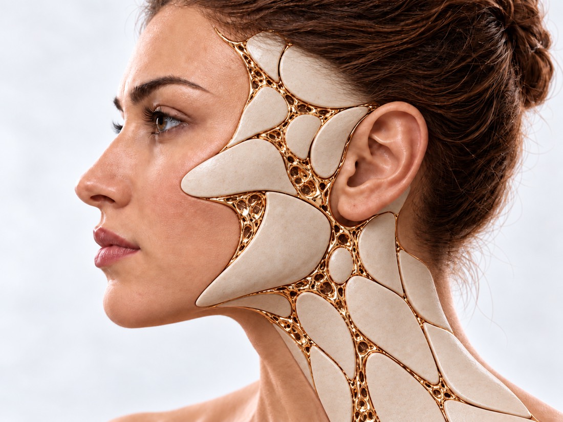
Conceptual visualization: reconstruction of eyelids and peri-ocular areaCONCEPTUAL VISUALIZATION
What it is
Any lumps, nodules and spots on the eyelids and around the eyes.
How often malignant
From 5% to a quarter of eyelid formations in different clinics; most often - basal cell carcinoma.
The main thing
An accurate diagnosis can only be given by histology — the examination of the removed tissue.
Who treats it
A surgeon who operates on the eyelids; with cancer - together with an oncologist.

What is
neoplasms of the eyelids.

Eyelid neoplasms are lumps, nodules, cysts, and spots on the skin and edges of the eyelids, in and around the corners of the eyes. They are divided into benign, which do not grow into neighboring tissues and do not spread throughout the body, and malignant - cancer.

Most formations are benign. In a large study of 4,521 eyelid tumors, 5.0% were malignant, in another clinic, 25.2%. Patients with malignant tumors are older on average: 72.5 years versus 55.4. The area around the eyes is a place where skin cancer occurs more often than in other areas of the body.

The formation can be confusing to the eye: even an experienced doctor cannot always distinguish a benign from a malignant one without histology. Therefore, the removed formation is examined under a microscope. And since the eyelid protects the eye, after its removal, it is restored so that it closes and does not pull.

This is general information. The doctor will tell you what will suit you after the examination.

What to turn to
attention

Most bumps on the eyelids are safe. But some signs are a reason to show the formation to a doctor and not to postpone its removal with histology.

01

Growing or changing

The formation gradually increases, changes color or shape, has uneven edges; small blood vessels are visible on it.

02

Eyelashes fell out

If the growth at the edge of the eyelid has shed eyelashes, in one study the odds of it being malignant were 13.4 times higher. But even without eyelash loss, cancer is possible.

03

Ulcer, crust, compaction

Central wound, induration, bleeding or crusting for more than 4 weeks. In one study, a cancer that was mistaken for a benign tumor to the eye mostly looked like a normal nodule with no visible changes on the surface.

Basal cell carcinoma of the face
04

"Halazion" that does not go away

A chalazion is an inflammatory "greenhouse" in the thickness of the eyelid. It can be disguised as sebaceous carcinoma — cancer of the eyelid glands. A chalazion that returns or does not go away is alarming, but cancer also occurs under the guise of the first chalazion.

Which are
neoplasms of the eyelids.

The type of formation depends on how widely it is removed and whether treatment by an oncologist is required. A well-known risk factor for basal cell carcinoma is strong ultraviolet radiation: skin cancer most often occurs on areas exposed to the sun, particularly on the face.

Benign

The most common are moles (nevi), papillomas, seborrheic keratomas, cysts, and xanthelasma. They are removed if they interfere, change or cause doubt.

Xanthelasma of the eyelids

Basal cell carcinoma

The most common malignant tumor of the eyelids is more than half of all malignant tumors in large series. More than 50% of eyelid basal cell carcinomas appear on the lower eyelid. It is advised to remove it without delay.

Sebaceous carcinoma

A rare but potentially aggressive cancer of the sebaceous glands of the eyelid; more often on the upper eyelid. It is often mistaken for a chalazion, and it can spread to the lymph nodes.

Squamous cell carcinoma and melanoma

Squamous cell carcinoma often resembles basal cell carcinoma and is treated similarly. Melanoma occurs less often, but is more dangerous - it often metastasizes.

Squamous cell carcinoma and melanoma

When to seek medical advice?

Neoplasms of the eyelids usually do not require emergency care, but a suspicious formation should not be delayed: the earlier the cancer is removed, the easier the treatment. After eyelid surgery, some symptoms require immediate help.

103 immediately if:

  • suddenly stopped seeing in one or both eyes;
  • sudden severe pain in the eye;
  • after surgery on the eyelid - severe pain in the eye or around it, which increases;
  • after eyelid surgery, the eye protrudes or became tense;
  • after eyelid surgery, the eyelids do not open;
  • double vision or impaired vision after eyelid surgery.

To the doctor, depending on the symptoms:

On the same day: the eyelid is swollen, red, hot and painful; the eyelid suddenly drooped; the eye does not open; vision has changed; the white of the eye is very red; high fever, chills or general malaise. In the coming weeks: the formation on the eyelid grows, changes color or shape, bleeds or is covered with a crust for more than 4 weeks; eyelashes fall out; "chalazion" returns or does not go away. Planned: an ancient hump that does not change, but interferes or disturbs.

As defined,
what kind of formation is this

  1. Conversation

    How long ago did the formation appear, is it growing, was something removed in this place; whether you or a family member has had skin cancer, whether you spend a lot of time in the sun.

  2. Examination of the eyelids

    Edges, color, eyelashes, ulceration or thickening, as a formation associated with the edge of the eyelid and corners of the eye.

  3. Histology

    The removed tissue or a piece of it (biopsy) is examined under a microscope - this is the only way to make an accurate diagnosis. In one study of 86 biopsies, in 4 cases (4.6%) a mass that was considered benign to the eye turned out to be malignant.

  4. Additional examinations

    For sebaceous carcinoma and large aggressive tumors, the oncologist may order a lymph node examination and imaging to determine the spread.

How we treat
neoplasms of the eyelids.

The main treatment of almost all formations of the eyelids is surgical removal. How to widely remove is decided by the type of formation; after removing the eyelid is restored.

01

Benign formations

Removal with histology
Formations are cut out and necessarily sent for histology.
Chalazion
If it is removed, the tissue is examined for both primary and recurrent chalazion. In a study of 1,060 "chalazions," 1.4% were malignant, mostly sebaceous carcinoma, and most were primary.
02

Malignant tumors

It is important to remove the tumor completely, with clean edges.

Edge control
The edges of the removed tissue are checked under a microscope even during the operation - excision with urgent examination of the edges or micrographic surgery, when the tumor is removed in layers and each layer is checked at once. After such methods, basal cell carcinoma around the eye returned in 1.9% and 2.9% of cases, after excision with planned histology — in 5.9%.
Choice of method
There are no randomized studies that would compare the methods with each other, so it is impossible to say unequivocally which of them has fewer relapses.
Treatment at an oncologist
In addition to surgery, radiation therapy and drugs (vismodegib, imiquimod) are used for basal cell carcinoma - they are prescribed by an oncologist depending on the type and spread of the tumor. In case of large or aggressive tumors, the lymph nodes are examined. If the tumor has grown into the eye socket, sometimes its contents have to be removed.
03

Eyelid reconstruction

The goal
Restore the shape and function of the eyelid by replacing the lost with similar tissues: the eyelid must protect the eye and close.
Large defects
Malignant tumors are removed with a reserve of healthy tissue, so the defect can be large - then more complex reconstructive operations on the eyelids are required.
04

Result and observation

Relapse
Malignant tumors of the eyelids return more often than benign: 11.9% versus 4.4% in a study of 4,521 formations. After micrographic surgery, recurrence was 3.0% at a median of 46 months.
After surgery
In a study of 32 patients with sebaceous carcinoma, the most common postoperative problem was eye irritation (30%). Regular check-ups are required.

How
we treat it.

Neoplasms of the eyelids at the Dr. Valikhnovskyi Institute of Surgery are treated in the "New Face" direction. The clinic is responsible for the removal of the mass with histology and for the reconstruction of the eyelid. If the tumor turns out to be malignant, further treatment and observation is conducted by an oncologist, and the condition of the eye is monitored by an ophthalmologist.

CLINIC SERVICE A new face Form, symmetry, facial expressions and functions after injuries, wounds, burns, removal of tumors and congenital changes. View service

What we do for this condition

  • Removal with histology — formations are cut out and sent for histological examination.
  • Reconstruction in the same operation — if the edges of the incision are clean, the eyelid is restored immediately; otherwise - after histology.
  • Local patches — the defect is covered with tissues from neighboring areas of the face, closest in color and texture.
  • Transplants and spacers — if there is a lack of tissue, a skin or cartilage graft or a spacer is used so that the eyelid has support.
  • Canthopexy and canthoplasty - strengthen or restore the corner of the eye so that the lower eyelid fits tightly to the eye.

Rostislav Valikhnovskyi

Plastic surgeon, candidate of medical sciences, founder of Dr. Valikhnovskyi Institute of Surgery.

How treatment works with us

  1. Photo onlineSend a close-up photo of the formation and the eyelids straight - the doctor will pre-evaluate the situation.
  2. ConsultationExamining the eyelids, finding out how suspicious the mass is, and planning for removal.
  3. RemovalThe formation is cut out, the tissue is sent for histology; the eyelid is restored immediately or after the result.
  4. The result of histologyThe doctor will explain the conclusion; if the tumor is malignant, he will refer to an oncologist for a further plan.
  5. RecoverySwelling of the first days, suture care and follow-up examinations to detect a relapse in time.

Common
questions

Short answers. What is right for you, the doctor will tell you during the consultation.

How to understand that the formation on the eyelid is malignant?

Warn growth, ulcers, compaction, uneven edges, blood vessels and eyelash loss. But only histology can give an exact answer.

Can a chalazion turn out to be cancer?

Rarely, but maybe. Sebaceous carcinoma is most often disguised as a chalazion. Therefore, the removed chalazion is examined - both the first and the second.

Why histology, if the doctor already sees what it is?

On the eye, the diagnosis does not always match the histology: in one study, in 84% of cases, and in 4 out of 86 cases (4.6%), the formation, which was considered benign, turned out to be malignant.

Is eyelid basal cell carcinoma dangerous?

This is the most common malignant tumor of the eyelids. It is advised to remove it without delay: if it grows into the eye socket, the treatment becomes much more difficult.

Is it possible to remove the tumor and restore the eyelid in one operation?

Often yes - if the edges of the cut are clean. If not, the eyelid is restored after the results of histology.

Can the tumor come back?

Maybe Malignant tumors of the eyelids return more often than benign tumors, so after treatment, follow-up examinations are required.

Do you need an oncologist and an ophthalmologist?

If the tumor is malignant, further treatment and observation is conducted by an oncologist. The condition of the eye and conjunctiva is assessed by an ophthalmologist.

Sources

18 sources · reviews, guidelines and research
showCollapse
  1. Comparison of the Clinical Characteristics and Outcome of Benign and Malignant Eyelid Tumors: An Analysis of 4521 Eyelid Tumors in a Tertiary Medical CenterBioMed Research International, 2015doi:10.1155/2015/453091 ↗
  2. Demographic features and histopathological diagnosis in primary eyelid tumors: results over 19 years from a tertiary center in Ankara, TurkeyInternational Journal of Ophthalmology, 2020doi:10.18240/ijo.2020.08.16 ↗
  3. Periocular skin cancer: Diagnosis and managementDisease-a-Month, 2020doi:10.1016/j.disamonth.2020.101046 ↗
  4. Ocular basal cell carcinoma: a brief literature review of clinical diagnosis and treatmentOncoTargets and Therapy, 2017doi:10.2147/OTT.S130371 ↗
  5. Eyelid lesions in general practiceAustralian Journal of General Practice, 2019doi:10.31128/AJGP-03-19-4875 ↗
  6. Madarosis as an indicator for malignancy in eyelid margin lesionsOptometry and Vision Science, 2012doi:10.1097/OPX.0b013e31824352b6 ↗
  7. Clinical Signs for Differential Diagnosis of Eyelid TumoursKlinische Monatsblätter für Augenheilkunde, 2023doi:10.1055/a-1996-0950 ↗
  8. Eyelid tumors: accuracy of clinical diagnosisAmerican Journal of Ophthalmology, 1999doi:10.1016/s0002-9394(99)00302-5 ↗
  9. Accuracy of the clinical diagnosis of chalazionEye, 2004doi:10.1038/sj.eye.6700603 ↗
  10. Sebaceous carcinoma of the eyelid: 21-year experience in a Nordic countryActa Ophthalmologica, 2021doi:10.1111/aos.14552 ↗
  11. Sebaceous Carcinoma: A Review of the Scientific LiteratureCurrent Treatment Options in Oncology, 2017doi:10.1007/s11864-017-0490-0 ↗
  12. Recurrence rates following surgical excision of periocular basal cell carcinomas: systematic review and meta-analysisJournal of Dermatological Treatment, 2020doi:10.1080/09546634.2019.1698702 ↗
  13. Mohs micrographic surgery versus surgical excision for periocular basal cell carcinomaCochrane Database of Systematic Reviews, 2014doi:10.1002/14651858.CD007041.pub4 ↗
  14. Recurrence of periocular basal cell carcinoma and squamous cell carcinoma after Mohs micrographic surgery: a retrospective cohort studyBritish Journal of Dermatology, 2019doi:10.1111/bjd.17516 ↗
  15. Eyelid ReconstructionPlastic and Reconstructive Surgery, 2017doi:10.1097/PRS.0000000000003820 ↗
  16. Eyelid problemsNHS, 2023nhs.uk/symptoms/eyelid-problems ↗
  17. Symptoms of non-melanoma skin cancerNHS, 2026nhs.uk/conditions/non-melanoma-skin-cancer/symptoms ↗
  18. Vision lossNHS, 2025nhs.uk/conditions/vision-loss ↗

The figures on the page are from these sources.

VALIKHNOVSKI SURGERY PARTNERS

Lump on the eyelid
bother?

Send a photo of the formation. The doctor will tell how suspicious it is, how to remove it and restore the eyelid.

Call Send an enquiry
ONLINE ENQUIRY

Tell us how we can help.

Leave your contact details and briefly describe the situation. A coordinator will contact you to agree on the next step.

Preferred contact method