Nothing bothers you
The fragment is visible only in the picture, but it does not hurt and does not interfere.
 Discuss your case
Shrapnel in the face and neck are fragments of shells, bullets, glass, wood, or other materials left in the skin and soft tissues after an injury. Not every fragment needs to be removed: the decision is made after examination and CT, weighing the benefits and risks of the operation.
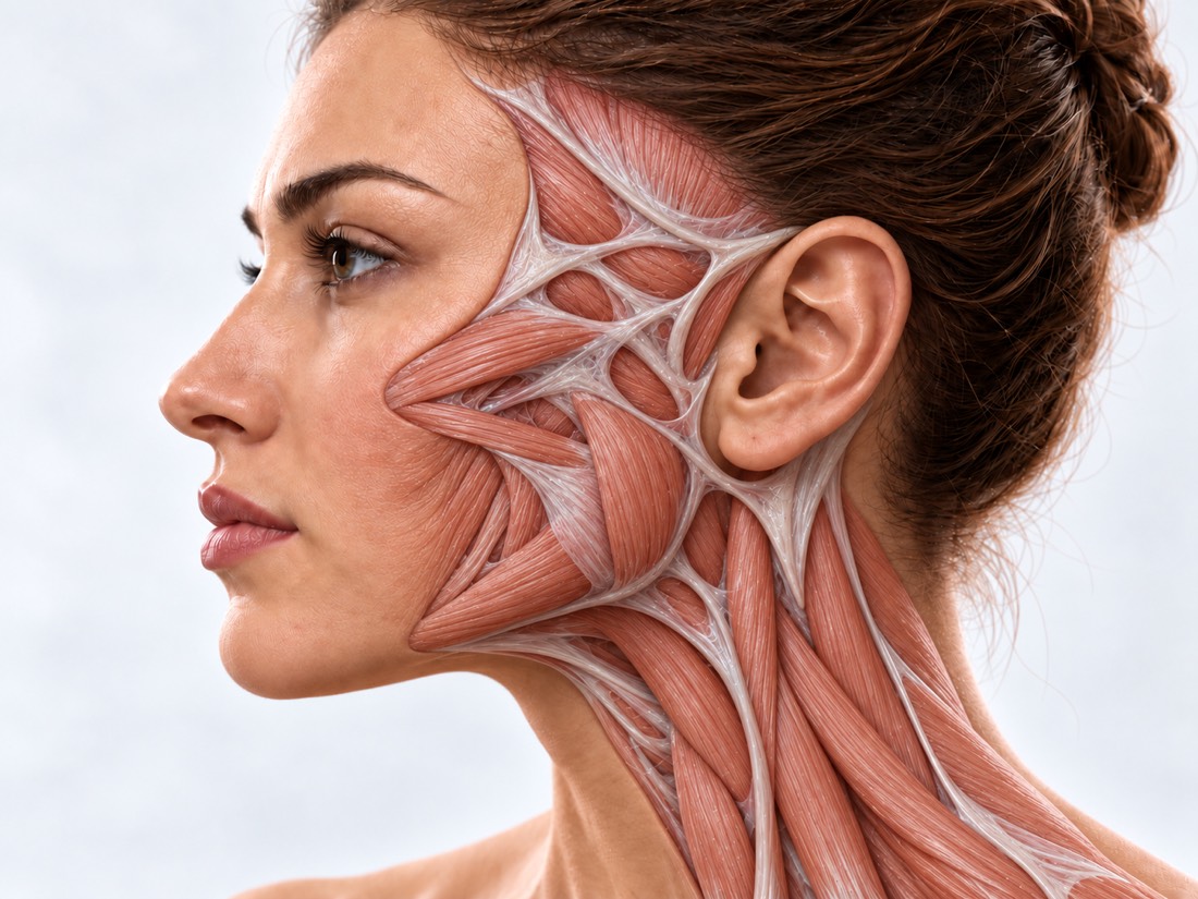 CONCEPTUAL VISUALIZATION
A foreign body is everything that entered the tissues from the outside and remained there. After injuries, these are fragments of shells and mines, parts of bullets, as well as glass, wood, pebbles or plastic, which are driven into the wound by an explosion or impact. Most often, glass, metal and wood are found in wounds.
The body tries to isolate the foreign object: inflammatory tissue forms around it. Part of the debris does not bother. Others cause infection—most often complications—long-term pain or pressure on nerves and blood vessels.
Therefore, the main question is not "how to get everything", but "what exactly should be deleted". On the face, a wide search for a fragment through a large incision is undesirable: facial expressions, nerves and appearance are important here.
This is general information. The doctor will tell you what will suit you after the examination.
Complications from fragments are early and late: the fragment may not bother you for a long time, and then catch fire.
The fragment is visible only in the picture, but it does not hurt and does not interfere.
It feels hard under the skin, hurts when touching, chewing or turning the head.
Redness, swelling, pus, a wound or scar that reopens. Infection is most often a complication of foreign bodies.
Fire woundFragment near the nerve: part of the face is numb or moves less.
Paralysis of the facial nervePain, bulging of the eye, new double or blurred vision. If it appeared suddenly or after a fresh injury, call 103; the fragment in the orbit is evaluated together with an ophthalmologist.
Injuries of the orbitAfter the injury, scars remained with the fragments under them; the scar tightens the skin or hurts.
Scars after facial injuriesThe material of the fragment depends on whether it is visible on the picture, how prone it is to infection and whether MRI is safe.
Fragments of shells, mines and drones. In a study of 53 wounded from the war in Ukraine, who were treated in Spain, 83% were affected by the explosion.
Bullet fragments may contain lead, and some military bullets may contain steel or nickel.
These fragments are often missed during examination and on a conventional X-ray; wood and plastic X-ray may not show at all.
In a study of war wounded in Ukraine, wound infection was associated with the presence of foreign bodies, bone damage, and skin defect.
An old wreck that does not bother you is a planned situation, it usually does not need emergency help. But a fresh wound on the neck or face can be dangerous: the neck is close to large blood vessels and airways.
On the same day: the skin around the fragment is reddened, swollen, hurts more and more, or pus flows from the wound; you feel unwell or have a fever; earth or dirt in the wound that does not wash out; the eye near the old fragment is reddened or vision is gradually deteriorating - see an ophthalmologist. If the wound is fresh, the doctor will also check the tetanus vaccination. Planned: the fragment is felt under the skin, hurts, interferes, or just bothers you - see a CT surgeon to decide whether to remove it. If bullet fragments remain in the body, ask your doctor about a blood test for lead.
How and when the wounding occurred, whether the fragments were already removed, which is troubling now; statements and photographs, if available.
The surgeon evaluates scars, induration, signs of inflammation, facial expressions and sensitivity.
Basic examination: shows metal fragments, their size and distance to vessels, nerves and the eye. Without accurate pictures, the debris is not removed.
Sees both metal and non-metal debris, if they lie shallow. According to a meta-analysis, ultrasound detects 72% of foreign bodies in soft tissues.
Fragments with steel in the magnetic field can shift and damage nearby tissues, and the composition of the fragment is unknown. Therefore, MRI for metal fragments is performed only after a risk assessment by a radiologist, and CT is used to search for fragments. Before any MRI, be sure to tell about the debris.
There are no single rules for which debris to remove. Decisions are made individually: some fragments can be left, but if the risk of complications is high, they are removed. The doctor discusses the benefits and risks of the operation with the patient.
When the debris damages or may cause damage.
When the operation is more risky than the wreck itself.
Splinters and foreign bodies in the soft tissues of the face and neck are treated at Dr. Valikhnovskyi's Institute of Surgery in the direction of "Surgery for gunshot wounds." The clinic decides whether to remove the fragment and removes it; if the fragment is in the eye, it is treated by an ophthalmologist.
Plastic surgeon, candidate of medical sciences, founder of Dr. Valikhnovskyi Institute of Surgery.
Short answers. What is right for you, the doctor will tell you during the consultation.
No. Debris that hurts, causes infection, lies next to the nerve, as well as wood and plant fragments are removed. Deep metal fragments without symptoms are sometimes left and observed. The decision — after examination and CT.
Only after a risk assessment by a radiologist: debris with steel in a magnetic field can shift, and the composition of the fragment is unknown. A CT scan is used to search for debris. Before any MRI, be sure to tell about the debris.
Lead poisoning from bullet fragments is rare, but can take months or years to manifest. If there are several fragments or there is a fracture, the doctor may prescribe a blood test for lead.
If the injury is fresh, call 103, even if the bleeding is almost nonexistent: the hospital will evaluate the condition with a CT scan with vascular contrast. If the fragment is old and does not bother you, the surgeon will tell you whether such a CT scan is necessary. Debris near vessels is removed only as indicated.
It depends on where the fragment lies. On the face, a wide search through a large incision is undesirable, so the fragment is first accurately found on CT, and access is chosen for each fragment separately.
The scar from the incision always remains, but most scars lighten over time. If there are already rough scars after the injury, the surgeon will tell if they can be corrected during the same operation.
See a surgeon the same day: infection is most often a complication of foreign bodies. Such a fragment is usually removed.
The figures on the page are from these sources.
Send photos, CT scans and statements. The doctor will tell if the fragment should be removed and how to do it.
Leave your contact details and briefly describe the situation. A coordinator will contact you to agree on the next step.