CONDITION / FACE

Osteoma of the frontal bone and skull.

Osteoma of the frontal bone is a benign tumor of bone tissue that grows slowly. It can be a firm bump on the forehead or a finding on a CT scan in the sinus. Often does not cause symptoms; remove those that grow, interfere or change the appearance.

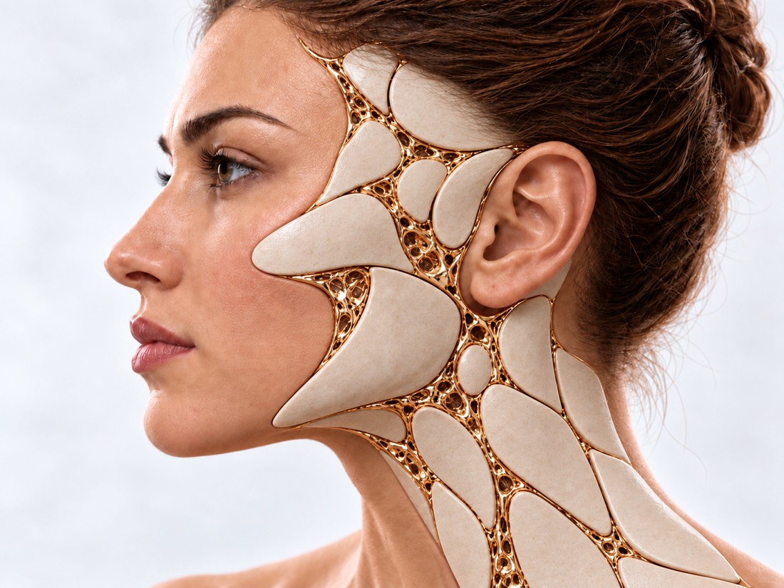
Conceptual visualization: smooth contour of the forehead and skull bonesCONCEPTUAL VISUALIZATION
What it is
Benign bone tumor that grows slowly; the most common benign tumor of the bones of the skull and face.
Where it happens
On the surface of the frontal bone there is a hump under the skin; or inside the sinuses — frontal, ethmoid.
How common
It was found on CT scans of the sinuses in 3% (1,889 examined) and 6.4% (1,724) of people who were examined due to suspicion of sinus disease.
Who treats it
Facial surgeon; with an osteoma of the sinuses — together with an ENT surgeon, near the eye — with an ophthalmologist.

What is
osteoma

Osteoma is a benign tumor of bone tissue that grows slowly. There is a central one - it grows from the inside of the bone, and a peripheral one - from the periosteum, a thin membrane on the surface of the bone.

Peripheral osteoma of the forehead is usually noticed by the person himself: a hard bump appears on the forehead. Forehead osteomas are mostly referred to plastic surgeons.

Osteomas inside the sinuses of the nose - frontal, ethmoid - usually do not cause symptoms and are accidentally found on a CT scan. Rarely, such an osteoma blocks the outflow of mucus from the sinus, presses on the eye or grows to the base of the skull - then complications arise.

This page is about the osteoma itself. There is a separate page about scars on the forehead called "Forehead cicatricial deformity", and about fractures - "Fractures of the frontal bone".

This is general information. The doctor will tell you what will suit you after the examination.

How
osteoma presents.

The osteoma of the forehead is visible from the outside. A sinus osteoma is usually silent until it becomes large or blocks sinus drainage.

01

A bump on the forehead

Firm and usually painless. In a study of 39 patients, the size of forehead osteomas ranged from 4 to 30 mm, with an average of 10 mm.

02

Discomfort

For the most part, an osteoma of the forehead does not bother you with anything, except for its appearance, but sometimes there is pain or discomfort in its area.

03

The sinuses

Congestion, pressure in the forehead area, repeated sinusitis. In a study of 1,889 people, 37% of sinus osteomas were accompanied by changes in the sinuses on CT.

Chronic frontal sinusitis and ethmoiditis
04

eyes

If the osteoma enters the orbit, the eye protrudes, doubles, the vision becomes blurred, the eyelids swell.

05

CT scan finding

Sinus osteoma is often found incidentally when a CT scan of the head or sinus is performed due to other complaints.

Why does it occur?
osteoma

The exact cause is unknown. But there are several patterns that help the doctor assess the situation.

There are more women among those operated on

In two surgical series, 82% (39 patients with forehead osteomas) and 79.1% (141 patients with craniofacial osteomas) were women. But the review of the literature does not find an advantage by gender.

Injury is rare

In a study of 39 patients with forehead osteoma, only two (5%) had a history of head trauma.

Frontal bone

In a series of 141 patients with osteomas of the skull and face with 148 osteomas on the frontal bone, there was 60.1%; then the parietal, lower jaw and occipital.

Benign tumors of the jaws

Smoking

In 47 patients, sinus osteomas in those who smoked or had smoked grew faster, though still slowly.

Gardner syndrome

A rare hereditary disease: multiple osteomas, extra or unerupted teeth and polyps of the intestine, which, without treatment, turn into cancer. Therefore, early diagnosis is important.

When to seek medical advice?

Osteoma grows slowly and usually does not require emergency care by itself. Dangerous rare complications of sinus osteomas — when they press on the eye or grow to the brain.

103 immediately if:

  • suddenly stopped seeing in one or both eyes;
  • severe pain in the eye suddenly appeared;
  • very high or very low body temperature, the person shivers;
  • very strong headache;
  • a person thinks confusedly or speaks unclearly;
  • convulsive attack for the first time in life;
  • the neck is stiff and it hurts to look at the light;
  • a rash on the skin that does not fade under a glass;
  • after an operation on the sinus or near the eye, pain in or around the eye increases;
  • after an operation on the sinus or near the eye, the eye protrudes or has become tense;
  • after an operation on the sinus or near the eye, the eyelids are swollen so that they do not open;
  • new diplopia has appeared or vision has worsened after surgery in the sinus or near the eye.

Do not delay:

On the same day - to the doctor: suddenly began to double in the eyes; the eye hurts and doubles; vision became blurred; swelling appeared above the eye or the eye began to protrude; a clear watery liquid constantly drips from the nose. In the coming days: the bump on the forehead has noticeably increased in a short time; congestion and pressure in the forehead area do not go away. Planned: a bump on the forehead that grows slowly; osteoma that was accidentally found on a CT scan - make an appointment for a consultation with images. If there are several osteomas, and there were polyps or intestinal cancer in the family, tell the doctor about it.

As defined,
what to do

  1. Review

    The doctor examines and feels the hump, asks when it appeared and whether it is growing, whether there are complaints from the nose and eyes.

  2. CT, in particular 3D

    The main examination: confirms that it is an osteoma, shows its size and connection with the sinuses, the eye, the base of the skull.

  3. MRI if needed

    If the sinus osteoma could affect soft tissues, the eye or the skull cavity.

  4. Ophthalmologist

    If there is an osteoma near the orbit or there are complaints about vision - a full eye examination.

  5. Multiple osteomas

    If there are several osteomas and they are large, genetic testing for Gardner syndrome may be recommended.

  6. Histology

    After removal, the tissue is examined under a microscope to confirm the diagnosis.

How we treat
osteoma

The operation is recommended when the osteoma gives symptoms or is actively growing, as well as when the person is concerned about the appearance of the forehead. Before operating, the risks of surgery are always weighed against the expected benefits — especially around the eye and the base of the skull.

01

Follow-up

Sinus osteoma without symptoms.

Slow growth
In 47 patients, sinus osteomas grew by an average of 0.42 mm per year. In another study (89 patients, mean follow-up of 54 months), osteoma increased in 46 of 89 patients.
Control CT scans
The authors of one study believe that part of the osteoma does not require observation, and if it can block the outflow from the sinus, one CT scan after a year is enough, and if the osteoma has increased, control images up to 3 years. Other authors advise monitoring, as serious complications are possible.
02

Osteoma of the forehead

A hump under the skin of the forehead.

Direct access
The osteoma is removed through an incision above it. In a study of 39 patients, 74% were operated on in this way, there was no regrowth in any of them.
Endoscopically
The osteoma is removed through a small incision along the hairline. In one series (9 patients) — through one incision of 3 cm in the middle, hidden 2 cm behind the hairline.
The result
In a series of 22 patients after endoscopic removal, swelling and bruising occurred in the first week and passed by themselves; there were no contour irregularities and no bone remains. The first three patients had partial baldness near the incision, after which the technique was changed.
Bone outline
After removal, the surface of the frontal bone is leveled, and the large defect is restored with its own bone or an implant.
03

Osteoma of the sinus

If it gives symptoms or complications.

Through the nose
Most frontal sinus osteomas, according to the authors of the review, can be removed endoscopically through the nose; it is limited only by osteomas that extend far forward or to the side. There was no regrowth in 34 patients operated on in this way.
Large osteomas
Osteomas over 3 cm or near the base of the skull and eye are more difficult to remove. In 160 such patients, part of the osteoma remained in 9.4%, complications were in 21.9%. Sometimes access through the nose and open is combined.
Near the eye
In 12 patients with osteoma extending into the orbit, after endoscopic surgery, ocular symptoms disappeared or significantly improved in more than 90%.

How
we treat it.

At Dr. Valikhnovskyi's Institute of Surgery, osteomas of the forehead and cranial vault are dealt with by the "New Face" department, where the forehead and cranial vault are a separate zone of reconstruction. The clinic is responsible for the bone and contour of the forehead; sinus osteomas requiring surgery through the nose and eye examination are conducted by an ENT surgeon and an ophthalmologist.

CLINIC SERVICE A new face Form, symmetry, facial expressions and functions after injuries, wounds, burns, removal of tumors and congenital changes. View service

What we do for this condition

  • CT and 3D planning — the size and boundaries of the osteoma are assessed by CT and contour restoration is planned.
  • Mirror CT modeling — the intact half is mirrored to reproduce the symmetrical contour of the forehead.
  • Contour of the forehead — a bone defect or unevenness is restored with one's own bone or an implant.
  • Individual implants — from medical titanium or PEEK according to the 3D model, if a bone defect remains after the removal of a large osteoma.
  • Lipofilling — own fat aligns the soft tissues over the bone after reconstruction.

Rostislav Valikhnovskyi

Plastic surgeon, candidate of medical sciences, founder of Dr. Valikhnovskyi Institute of Surgery.

How treatment works with us

  1. Photos and pictures onlineSend a photo of the forehead and a CT scan, if you already have one - the doctor will pre-evaluate the situation.
  2. ConsultationExamination, if necessary — CT with 3D reconstruction and consultations of an ENT doctor or an ophthalmologist.
  3. PlanObserve or delete; with what access and whether the contour of the bone needs to be restored.
  4. TreatmentRemoval of the osteoma and alignment of the forehead contour — or a control CT according to the plan.
  5. Follow-upInspection; histology confirms the diagnosis.

Common
questions

Short answers. What is right for you, the doctor will tell you during the consultation.

Is osteoma cancer?

No. This is a benign tumor of mature bone tissue that grows slowly.

Is it necessary to remove it?

No. Osteoma without symptoms, which does not grow, is often only observed. Surgery is advised if it causes symptoms, actively grows or changes the appearance of the forehead.

How fast does an osteoma grow?

slowly In one study of 47 patients, sinus osteomas grew by an average of 0.42 mm per year, in another (89 patients) by less than 1 mm per year.

Will there be a scar on the forehead?

A trace of the incision always remains. During endoscopic removal, the incision is made along the line of hair growth, so it is hidden; direct incision — above the osteoma.

Can the osteoma come back?

In studies of osteomas of the forehead (39 patients) and sinuses (34 operated on), there was no regrowth. It is not always possible to completely remove large osteomas of the sinuses: in 160 patients, a part remained in 9.4%.

Why does the doctor ask about the intestine?

Multiple osteomas occur in Gardner's syndrome, a hereditary disease with intestinal polyps that turn into cancer without treatment. If there is suspicion, the doctor will recommend an examination.

Is it possible to remove a sinus osteoma without an incision on the face?

This is often the case: many osteomas of the frontal sinus are removed endoscopically through the nose. Access is chosen by CT.

Sources

18 sources · reviews, multicentre and retrospective studies, NHS
showCollapse
  1. Frontal peripheral osteomas: a retrospective studyArchives of Craniofacial Surgery, 2023doi:10.7181/acfs.2022.01004 ↗
  2. Diagnostic and Treatment Protocols for Peripheral Craniofacial OsteomasJournal of Craniofacial Surgery, 2026doi:10.1097/SCS.0000000000012685 ↗
  3. Endoscopic-Assisted Excision of Frontal Bone Osteomas: A Case Series Study of Technical Considerations and Complications from a Single SurgeonMedicina, 2026doi:10.3390/medicina62050947 ↗
  4. Endoscopic management of osteomas of the foreheadBritish Journal of Oral and Maxillofacial Surgery, 2015doi:10.1016/j.bjoms.2015.06.004 ↗
  5. Osteomas of the craniofacial region: A case series and review of literatureIndian Journal of Plastic Surgery, 2013doi:10.4103/0970-0358.121982 ↗
  6. A prospective study of paranasal sinus osteomas in 1,889 cases: changing patterns of localizationThe Laryngoscope, 2009doi:10.1002/lary.20646 ↗
  7. Characteristics of paranasal sinus osteoma and treatment outcomesActa Oto-Laryngologica, 2015doi:10.3109/00016489.2014.1003093 ↗
  8. Growth characteristics and clinical manifestations of the paranasal sinus osteomasOtolaryngology–Head and Neck Surgery, 2011doi:10.1177/0194599811403380 ↗
  9. Growth rate, impact of smoking and management of incidentally found paranasal sinus osteomasAmerican Journal of Otolaryngology, 2024doi:10.1016/j.amjoto.2024.104427 ↗
  10. Osteoma of the skull base and sinusesOtolaryngologic Clinics of North America, 2011doi:10.1016/j.otc.2011.06.008 ↗
  11. Benign bony lesions of paranasal sinuses and skull base: from osteoma to fibrous dysplasiaCurrent Opinion in Otolaryngology & Head & Neck Surgery, 2024doi:10.1097/MOO.0000000000000955 ↗
  12. Advanced paranasal sinuses osteomas: a retrospective multicentric analysis on surgical management outcomes and intra- and postsurgical complicationsRhinology, 2025doi:10.4193/Rhin24.165 ↗
  13. Unusual Manifestations of Sinonasal Osteomas: A Narrative ReviewClinics and Practice, 2026doi:10.3390/clinpract16070126 ↗
  14. Outcomes of endoscopic sinus surgery (ESS) on sino-orbital osteomas: ophthalmic manifestations and visual outcomes from a 12-year multicenter experience and literature reviewBMC Ophthalmology, 2026doi:10.1186/s12886-026-05411-6 ↗
  15. Oral and Maxillofacial Manifestations of Gardner Syndrome: A Literature AnalysisJournal of Craniofacial Surgery, 2026doi:10.1097/SCS.0000000000011639 ↗
  16. Double visionNHS, 2024nhs.uk/symptoms/double-vision ↗
  17. Vision lossNHS, 2025nhs.uk/conditions/vision-loss ↗
  18. MeningitisNHS, 2026nhs.uk/conditions/meningitis ↗

The figures on the page are from these sources.

VALIKHNOVSKI SURGERY PARTNERS

A bump on the forehead
or osteoma on CT?

Please send forehead photo and CT scan if available. The doctor will tell if it is worth observing or if it is better to remove.

Call Send an enquiry
ONLINE ENQUIRY

Tell us how we can help.

Leave your contact details and briefly describe the situation. A coordinator will contact you to agree on the next step.

Preferred contact method