CONDITION / JAWS, MOUTH AND NECK

Fracture of the upper jaw.

A fracture of the upper jaw is an injury to the middle part of the face, after which the upper jaw, together with the teeth, is partially or completely separated from the skull. One of its signs is that the teeth do not close as before; treatment is aimed at returning the jaw to its place and restoring the bite.

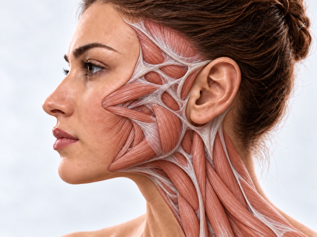
Conceptual visualization: restoration of the upper jaw and bite after a fractureCONCEPTUAL VISUALIZATION
What it is
A fracture, after which the upper jaw with teeth partially or completely separates from the skull.
How common
In the American surgical database, there are 130 such fractures out of 3,293 facial fractures.
The main risk
Immediately - breathing, bleeding, head and eye injuries; later - a changed bite.
Who treats it
A surgeon who operates on the bones of the face, together with a neurosurgeon, an ophthalmologist and a dentist.

What is
fracture of the upper jaw.

The upper jaw is the base of the middle part of the face: it houses the upper teeth, it forms the palate and the bottom of the nose. Doctors describe her fractures according to three typical Le Faure lines: type I — horizontal, type II — pyramidal, type III — transverse. All three separate the upper jaw from the skull, and the higher the line goes, the more part of the face is separated.

Such a fracture is the result of a strong blow to the middle of the face: most often a road accident, beating or fall, often under the influence of alcohol or drugs. In the American NSQIP surgical database for 2016–2019, 130 Le Fort fractures were found: 70 — type I, 41 — type II, 19 — type III. There were 4.9 men for every woman.

Mortality from Le Fort fractures alone is low, but they are rarely the only injury and are associated with other severe head and neck injuries. That is why they first rule out life-threatening conditions, and only then treat the bones.

This is general information. The doctor will tell you what will suit you after the examination.

How
fracture of the upper jaw presents.

Swelling in the first days can hide some of the changes, so the bite and the position of the bones are checked again when it subsides.

01

The teeth close differently

The upper teeth do not stand on the lower ones as before the injury. Le Faure fractures belong to those that disrupt the bite.

02

The jaw wobbles

Separated from the skull, the upper jaw can move: the upper teeth and the palate move as one.

03

Swelling, bruising, bleeding

The face swells greatly, bruises appear around the eyes, blood comes from the nose or mouth, it is difficult to breathe through the nose.

Fresh broken nose
04

Numbness

The cheek, upper lip or upper teeth do not feel the touch well - the fracture affected the nerve under the eye.

05

eyes

Double vision, pain in the eye, impaired vision - call 103. In a study of 106 patients with midfacial fractures, 41 had an eye injury, and 12 had an eye problem.

Fractures of the orbit
06

Cleft palate

Sometimes the fracture also runs along the palate — this is a sagittal fracture. In a series of 60 such patients, it was more often combined with Le Fort I and II fractures.

Why does it occur?
and what happens nearby.

It takes a lot of force to break the upper jaw, so there are often other injuries nearby.

A strong blow

Road accident, beating, fall. According to a review of 51 articles, such injuries are often associated with alcohol or drug use.

Other facial fractures

Fractures of the middle zone of the face can affect not only the upper jaw, but also the cheekbone, nose and orbit.

Fractures of the cheekbones

Lower jaw

In a series of 43 patients with a Le Fort I fracture, postoperative malocclusion was associated with a concomitant mandibular condylar fracture.

Fractures of the condylar process

Children

In small children, especially with baby teeth, the middle part of the face is rarely broken: the forehead protrudes more than it. Among 1,274 children with facial fractures, Le Fort fractures had 5.4%; they were associated with traffic accidents, older age, and brain injury.

When to seek medical advice?

Suspicion of a fracture of the upper jaw is a reason to see a doctor the same day: such a fracture occurs from a strong blow, and there may be an injury to the head, eyes, or neck. Do not get behind the wheel yourself. Call 103 if there is at least one of the signs:

103 immediately if:

  • it is difficult to breathe;
  • swelling in the mouth or neck grows rapidly, it is difficult to swallow;
  • bleeding from the nose or mouth does not stop;
  • a person has lost consciousness and does not regain consciousness;
  • the person is very sleepy, cannot keep his eyes open;
  • convulsions;
  • a clear, water-like liquid flows from the nose or ear;
  • double vision;
  • vision has worsened or disappeared;
  • impaired hearing;
  • the eye does not move, protrudes forward or hurts a lot;
  • pain in or around the eye increases after surgery;
  • bruises around the eyes, although there was no blow to the eye;
  • bleeding from the ear or bruising behind the ear;
  • neck pain with numbness or tingling in the hands;
  • new weakness or numbness in an arm or leg;
  • speech, gait or balance are disturbed, the person does not understand what is being said;
  • behavior has changed: irritability, inattention, indifference;
  • injury in a road accident or other impact at high speed;
  • falling from a height of more than 1 meter or from more than 5 steps;
  • a large wound on the face or glass or debris in the wound.

Do not delay:

Immediately to the reception department (let them take you away): after the impact, there is vomiting, dizziness in the head, a headache that does not go away, you lost consciousness for a short time, you are taking blood-thinning drugs, or the injury occurred under the influence of alcohol. On the same day - to the waiting room or to a surgeon who treats facial fractures: the teeth do not close as before after the impact; upper teeth wobble; numb cheek, upper lip or teeth; due to swelling, he cannot breathe through his nose. Hold the knocked-out tooth by the crown, without touching the root, and go to the dentist as soon as possible; if it is not possible to put it in place - carry it in milk or saliva. Don't worry. After surgery: pain or swelling of the face increases without signs from list 103 - immediately to the surgeon who operated, and if it fails - to the waiting room; high fever, chills or pus from the wound - the same day to the surgeon. In the coming days: if the fracture is treated without surgery — a control examination on the day appointed by the doctor. Planned: the fracture has grown, but the bite has changed or the face has become asymmetrical - make an appointment for a consultation.

How we make the
diagnosis

  1. Urgent review

    First, breathing, bleeding, consciousness, and the neck: concomitant injuries to the head and neck are more dangerous than the fracture itself.

  2. Bite and mobility

    The doctor looks at how the teeth close, whether the upper jaw moves, whether the cheek and upper lip feel the touch.

  3. CT of the facial skull

    Computed tomography with three-dimensional reconstruction shows the type of fracture according to Le Faure, displacement of fragments and fractures of the palate, cheekbones, orbit.

  4. eyes

    An ophthalmologist's examination is required already during first aid: if vision is at risk, the time to save it is short.

  5. Blood vessels and liquor

    After a severe stroke, CT angiography of the vessels of the neck is sometimes performed: their damage is rare (0.45% according to a systematic review), but can cause a stroke. Clear liquid from the nose is checked for cerebrospinal fluid.

How we treat
fracture of the upper jaw.

There are few high-evidence studies on the treatment of Le Fort fractures, so the plan is based on the type of fracture, displacement of fragments, and bite. The main thing is to turn the jaw so that the teeth close, as before the injury.

01

Without surgery

Fractures without displacement.

Follow-up
In children, fractures without displacement are usually only observed. In a cohort of 218 children, three-quarters of midfacial fractures were treated without surgery, with a low incidence of complications.
The first days
Soft food according to the doctor's advice; do not pick or pick your nose until the fracture heals.
02

Surgery

Displaced fragments, changed bite.

Comparison by bite
The fragments are returned to their place, focusing on how the teeth close, and fixed with plates. Accurate occlusal alignment is crucial for a good result.
Intermaxillary fixation
In order to maintain a correct bite, the jaws can be temporarily connected to each other - this is how, in particular, patients were treated in a series of 60 cases of fracture of the palate.
Palate
In some types of cleft palate with displacement, it is fixed with a plate. In a series of 60 patients, the most frequent complications were incorrect bite, exit of the plate through the mucosa, and connection between the mouth and the sinus — 2 cases each.
Risks
In the NSQIP database (130 patients), 5.4% of patients had in-hospital complications (infection or wound dehiscence, sepsis), reoperation — 2.3%. In a series of 43 patients after surgery for a Le Fort I fracture, malocclusion was present in 6, sensory disturbances in 4, and facial deformity in 6.
Children
Displaced fractures in children are compared and fixed, and then the growth of the face is monitored for a long time.
03

Concomitant injuries

What is treated along with the fracture.

Liquorice
Among 1,473 patients with jaw and facial trauma, 4.5% had CSF leakage that did not resolve after 5 days of treatment without surgery; in 84.9% of them it stopped after fracture treatment.
eyes
Eye injuries are treated by an ophthalmologist; bone surgery is planned taking into account the condition of the eye.
04

If it grew incorrectly

Months and years after the injury.

Repeat reconstruction
Even after a careful first operation, some patients need a second operation to restore facial shape and function.
Osteotomy
If the jaw has grown with a displacement and the bite is disturbed, in difficult cases it can be dissected along the Le Fort I line and rearranged (osteotomy).
Virtual planning
Asymmetry of the face can be corrected or significantly reduced if the healthy half of the face is mirrored on the damaged one and the operation is planned on the computer.

How
we treat it.

At Dr. Valikhnovskyi's Institute of Surgery, fractures of the upper jaw and their consequences are dealt with by the "New Face" department, where the cheekbone, orbit, and lower jaw are also treated. The clinic is responsible for facial bones; brain trauma is managed by a neurosurgeon, eyes by an ophthalmologist, teeth by a dentist.

CLINIC SERVICE A new face Form, symmetry, facial expressions and functions after injuries, wounds, burns, removal of tumors and congenital changes. View service

What we do for this condition

  • CT decision — whether an operation is necessary is decided based on the displacement of fragments, symptoms and CT scan; some fractures are treated without surgery or with closed reduction.
  • Titanium plates — displaced fragments of the upper jaw are compared and fixed.
  • Osteotomy — the jaw, which has grown together with displacement, is dissected and rearranged.
  • Mirror CT modeling — the intact half of the face is mirrored and a three-dimensional restoration plan is obtained.
  • Individual implants and templates — plates made of titanium or PEEK according to the 3D model; printed templates help to make cuts at a given angle.
  • Fibula flap — if a part of the upper jaw is lost, it is reproduced with a leg bone with its own vessels according to indications.

Rostislav Valikhnovskyi

Plastic surgeon, candidate of medical sciences, founder of Dr. Valikhnovskyi Institute of Surgery.

How treatment works with us

  1. Photo and CT scan onlineShow a photo of the face before the injury and the current bite, CT images and discharges - the doctor will assess the situation beforehand.
  2. ConsultationExamination of the bite, mobility of the jaw, sensitivity of the cheek and eyes; if necessary, an examination by an ophthalmologist or dentist.
  3. PlanIs surgery necessary and how to match the fragments by bite; for long-standing fractures — virtual CT planning.
  4. SurgeryAlignment and fixation of fragments with plates or osteotomy, if the jaw has already fused incorrectly.
  5. RecoverySome time the food is only soft or liquid. Do not exercise until the surgeon allows it, and do not smoke while the healing process is ongoing.

Common
questions

Short answers. What is right for you, the doctor will tell you during the consultation.

Is a fracture of the upper jaw always operated on?

No. Some fractures are treated without surgery — in particular, non-displaced fractures in children are usually only observed. If the fragments are displaced and the bite has changed, they are compared and fixed.

Why does the doctor look so carefully at the bite?

The upper teeth are held on the upper jaw. If it is not correctly matched, the teeth will not close as before the injury, so the fragments are returned to their place precisely by the bite.

The fracture has grown, but the teeth do not close properly. What to do?

Sign up for a consultation. The jaw, which has grown together with displacement, can be dissected and rearranged (osteotomy); the operation is planned for CT.

After a blow, a clear liquid flows from the nose. How is it dangerous?

This is how cerebrospinal fluid can leak out, and this is a sign of a severe head injury. Call 103 immediately.

Why can't you move after a fracture?

This is advised both for a broken nose and after operations on the upper jaw. Do not try until the doctor allows.

What to eat after surgery?

Some time - only soft or liquid food. Exactly how much, the surgeon will say.

A tooth was knocked out during the impact. What to do?

Hold the tooth by the crown, without touching the root, and go to the dentist as soon as possible; if it is not possible to put it in place - carry it in milk or saliva. If there are symptoms from the 103 list, call 103 first.

Sources

21 sources · reviews, databases and patient series, NHS and NICE
showCollapse
  1. Le Fort Fractures: A Collective ReviewBulletin of Emergency and Trauma, 2017doi:10.18869/acadpub.beat.5.4.499 ↗
  2. Le Fort FracturesStatPearls, 2024pubmed.ncbi.nlm.nih.gov/30252316 ↗
  3. LeFort and Complex Midface FracturesFacial Plastic Surgery Clinics of North America, 2025doi:10.1016/j.fsc.2025.03.007 ↗
  4. Age-Related Outcomes in Le Fort FracturesThe American Surgeon, 2023doi:10.1177/00031348231161683 ↗
  5. Management of Le Fort I FracturesCraniomaxillofacial Trauma & Reconstruction, 2024doi:10.1177/19433875241278796 ↗
  6. Sagittal Maxillary Fracture: Diagnosis and ManagementIndian Journal of Plastic Surgery, 2021doi:10.1055/s-0041-1729665 ↗
  7. The Epidemiology and Management of Pediatric Maxillary FracturesJournal of Craniofacial Surgery, 2021doi:10.1097/SCS.0000000000007050 ↗
  8. Pediatric Midface Fractures: Outcomes and Complications of 218 PatientsLaryngoscope Investigative Otolaryngology, 2019doi:10.1002/lio2.315 ↗
  9. Pediatric Le Fort, Zygomatic, and Naso-Orbito-Ethmoid FracturesOral and Maxillofacial Surgery Clinics of North America, 2023doi:10.1016/j.coms.2023.04.004 ↗
  10. Association of Midfacial Fractures with Ophthalmic InjuryCraniomaxillofacial Trauma & Reconstruction, 2017doi:10.1055/s-0037-1599228 ↗
  11. Maxillofacial trauma and cerebrospinal fluid leak: a retrospective clinical studyAfrican Health Sciences, 2023doi:10.4314/ahs.v23i4.41 ↗
  12. Blunt cerebrovascular injury following craniomaxillofacial fractures: A systematic reviewThe Laryngoscope, 2017doi:10.1002/lary.26186 ↗
  13. Emergency Department Management of Maxillofacial TraumaEmergency Medicine Practice, 2024pubmed.ncbi.nlm.nih.gov/39353207 ↗
  14. Secondary reconstruction of maxillofacial traumaCurrent Opinion in Otolaryngology & Head & Neck Surgery, 2017doi:10.1097/MOO.0000000000000368 ↗
  15. Integrating Virtual Planning and three-dimensional Printing for Craniofacial Trauma ManagementOral and Maxillofacial Surgery Clinics of North America, 2025doi:10.1016/j.coms.2025.04.009 ↗
  16. Head injury and concussionNHS, 2025nhs.uk/conditions/head-injury-and-concussion ↗
  17. Broken noseNHS, 2023nhs.uk/conditions/broken-nose ↗
  18. NosebleedNHS, 2023nhs.uk/conditions/nosebleed ↗
  19. Black eyeNHS, 2023nhs.uk/conditions/black-eye ↗
  20. Knocked-out toothNHS, 2025nhs.uk/conditions/knocked-out-tooth ↗
  21. Head injury: assessment and early management (NG232)NICE, 2023nice.org.uk/guidance/ng232/chapter/Recommendations ↗

The figures on the page are from these sources.

VALIKHNOVSKI SURGERY PARTNERS

After the impact
do the teeth close differently?

Send a CT scan and a photo: how the teeth close now and what the face was like before the injury. The doctor will tell you whether an operation is necessary.

Call Send an enquiry
ONLINE ENQUIRY

Tell us how we can help.

Leave your contact details and briefly describe the situation. A coordinator will contact you to agree on the next step.

Preferred contact method