CONDITION / FACE

Fracture of the cheekbone and the cheekbone.

A cheekbone fracture is an injury to the bone that forms the protrusion of the cheek and part of the eye socket, or the thin cheekbone on the side of the face. After it, the cheek may become flatter, numb, and the mouth may be harder to open; some fractures are treated without surgery, and the displaced bone is returned to its place and fixed.

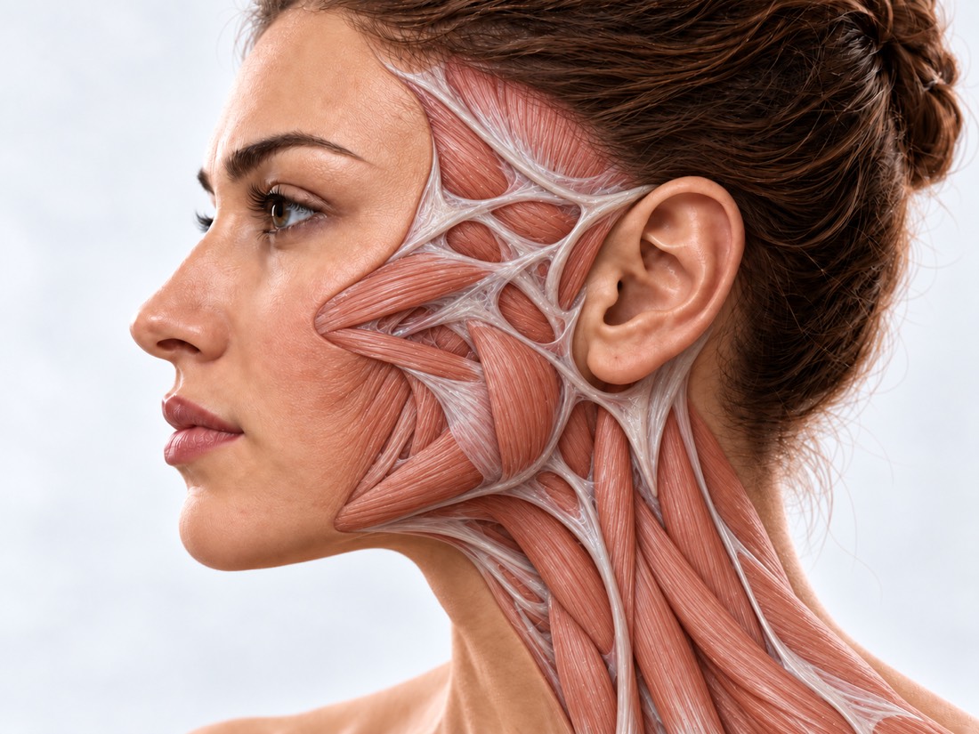
Conceptual visualization: reconstruction of the cheekbone and midface areaCONCEPTUAL VISUALIZATION
What it is
Fracture of the "rest" of the cheek or thin arch on the side of the face after a blow or fall.
How to notice
The cheek is flatter than the other, the cheek or upper lip is numb, it is difficult to open the mouth.
When surgery is performed
When the bone is noticeably displaced or functions are impaired; some fractures are treated without surgery.
Who treats it
A surgeon who operates on the bones of the face; if the eye is injured, together with an ophthalmologist.

What is
cheekbone fracture.

The cheekbone is the "support" of the cheek. It is connected to the neighboring bones of the skull and forms part of the bottom and side wall of the eye socket.

Doctors distinguish between two main options. Fracture of the facial complex — when the bone breaks at several points of connection and is displaced along with the edge of the eye socket. Isolated fracture of the facial arch - when only a thin bone "bridge" on the side of the face in front of the ear collapses.

Fractures of the facial complex are the second most common facial fractures after nasal fractures. A displaced cheekbone makes the middle part of the face flatter and wider, and due to a change in the volume of the eye socket, the position of the eye can change.

This is general information. The doctor will tell you what will suit you after the examination and CT scan.

How
cheekbone fracture presents.

In the first days, swelling and bruising can mask the flattening of the cheek, so the doctor evaluates not only the appearance, but also sensitivity, jaw and eye movements.

01

Flat cheek

The cheek on the side of the blow became flatter, the face became wider or asymmetrical.

02

Numbness

The touch of the cheek, the side of the nose, the upper lip or the upper teeth is difficult to feel - the nerve coming out under the eye has been affected.

03

It is difficult to open the mouth

The mouth does not open completely - a displaced bone can interfere with the movement of the lower jaw.

05

A step under the eye

A step is felt on the lower edge of the eye socket or on the cheek under the fingers, the place hurts when pressed.

06

Double or inflamed eye

If the fracture affects the bottom of the eye socket, the eye may double, move poorly, or sit lower. New diplopia or an unmoving eye after a stroke is a reason to call 103.

Orbital fracture

Why does it break?
cheekbone

The cause of the injury depends on what still needs to be checked, and the symptoms and treatment depend on how the fracture line passed.

Attack and fall

In a European study of 3,396 patients with primary facial fractures in different clinics, alternating attacks and falls; men predominated.

Road accidents and sports

Fractures of the cheekbone and arch are a frequent sports injury; they also happen in road accidents.

A line through the nerve canal

Numbness of the cheek depends not so much on displacement as on whether the fracture line passed through the nerve canal under the eye: then in a study of 272 patients, the chances of numbness were 2.7 times higher.

Incorrect splicing

If the fracture was not treated, was treated late, or the bone was not completely returned to its place, the cheekbone grows with displacement.

Post-traumatic deformations of the face

When to seek medical advice?

Suspected fracture of the cheekbone is a reason to see a doctor on the same day: a blow that breaks the cheekbone can affect the eye and head. If you hit your head, don't drive to the hospital. Call 103 if there is at least one of the signs:

103 immediately if:

  • difficulty breathing or swallowing;
  • swelling in the mouth or neck grows rapidly;
  • bleeding from the nose, mouth or wound does not stop;
  • the person fainted and does not regain consciousness;
  • a person cannot keep his eyes open, falls asleep all the time;
  • convulsions;
  • a clear watery liquid flows from the nose or ear;
  • bleeding from the ear or bruising behind the ear;
  • double vision appeared;
  • vision has worsened or disappeared;
  • impaired hearing;
  • the eye does not move;
  • severe pain in or around the eye or increasing pain;
  • the eye hurts when you move it;
  • the eye protrudes forward or has become tense and hard;
  • swollen eyelids do not open;
  • new numbness or weakness in an arm or leg;
  • impaired speech, understanding, writing, gait or balance;
  • pain or stiffness in the neck, and numbness or tingling in the hands;
  • a large wound on the face or glass or something else stuck in the wound;
  • a dent appeared on the head;
  • impact at high speed — for example, in an accident;
  • falling from a height of more than 1 meter or from five steps.

Do not delay:

Immediately - to the reception department (let them take you away): you can see blood in the eye; the pupil has lost its round shape; flashes, light circles or shadows in front of the eye; bright light hurts the eye; after a blow to the head - bruises under both eyes, vomiting or a person briefly lost consciousness. On the same day, to the surgeon who treats facial fractures, or to the waiting room: the cheek has become flatter or there is a step under the eye; numbness of the cheek, side of the nose, upper lip or teeth; it is difficult to open the mouth or the teeth close differently; the eye sits deeper or lower than the other; the headache does not go away. Likewise, on the same day, if you feel dizzy after hitting your head, you are taking blood thinners, or you drank alcohol before the injury. Do not rush to the review. After surgery: the pain in or around the eye increases, the eye protrudes or the vision worsens - this is 103. If the pain or swelling of the cheek increases without these signs - immediately to the surgeon who operated, and if it does not work - to the waiting room. If the temperature is 38 °C or higher or pus comes out of the wound, go to the surgeon on the same day. In the coming days: without surgery - a control examination when the doctor appoints. Planned: the fracture has long since healed, but the cheek remains flat, the face is asymmetrical or the eye is sunken, make an appointment for a consultation.

As defined,
whether surgery is necessary.

  1. Discussion and review

    How the blow happened; the symmetry of the cheeks, the step along the edge of the eye socket, the sensitivity of the cheek and lips, how the mouth opens and the teeth close.

  2. Eye examination

    Vision, movements and position of the eye. If there is diplopia, movement restriction or the eye is displaced, an ophthalmologist examines it.

  3. CT of the facial skull

    It shows how many joints are broken, how much the bone is displaced, whether the floor of the eye socket and the nerve canal under the eye are affected.

  4. Photo before the injury

    They show what the face looked like before the impact.

How we treat
cheekbone fracture.

The operation is recommended when the bone is noticeably displaced or the functions are disturbed - opening the mouth, vision, eye movements. Access and fixation are selected for a specific fracture.

01

Without surgery

If the debris is almost not displaced.

Who suits?
In a study of 103 patients with cheekbone fractures who were treated without surgery, incomplete fractures of one area of the bone were most suitable for this. For fragment fractures with significant displacement, the authors recommend surgery.
What passes and what does not
At 6 months, patients with less complex fractures had significant reductions in pain, limitation of mouth opening, and numbness, but flattening of the cheek, step under the eye, and changes on the side of the eye did not.
The first days
The first 1–2 days — ice wrapped in a cloth for 10–20 minutes several times a day. Do not press on the cheek or pinch; aspirin — only on doctor's prescription.
02

Arc control

When only the facial arch falls.

why
A sunken arch changes the contour of the face from the side and can make it difficult to open the mouth.
How
The arch is lifted into place with an instrument through a small incision or puncture of the skin. In a series of 15 patients in whom the arc was performed through a puncture, one had a complication—a hematoma—and two required conventional surgery through a temporal incision.
03

Repositioning and fixation

When the entire facial complex is displaced.

How
The cheekbone is returned to its place and fixed with small titanium plates in one, two or three points of connection with the neighboring bones. Incisions are made in the mouth, near the eyebrow or on the lower eyelid.
How many points
In a meta-analysis of 8 randomized trials (376 participants), fixation in three points could provide more stability than in two; enophthalmos and cheek asymmetry did not differ. Another review of 17 studies did not yield clear conclusions.
The bottom of the eye socket
In a series of 50 patients, the bottom of the orbit was restored only in the case of severe diplopia, restriction of movement or displacement of the eye; in 41, who were not opened, there were no signs from the side of the eye after the operation.
Eyelid
An incision on the lower eyelid can cause an inversion or inversion of the eyelid. In a meta-analysis of 17 studies (2,086 patients), the risk of inversion was highest after an incision under the eyelashes — 14%, inversion — after an incision on the inside of the eyelid, 1.5%; most of these complications passed without surgery.
Accuracy
For complex fractures, virtual planning, navigation and CT can help during surgery; there is still little evidence of their benefit.
04

Cheek numbness

What happens to him.

How common
According to a meta-analysis of 11 studies (802 patients), numbness in the area of the nerve under the eye after a fracture of the facial complex occurs in 51.9%. In a separate study of 272 patients — in 37.3%.
Recovery
In a study of 21 patients after surgery, the majority had markedly improved sensitivity at 3 months and almost completely recovered by 6 months.
pain
Long-term nerve pain in the cheek developed in 2.2% of 272 patients in one study.
05

Late correction

If the cheekbone has grown incorrectly.

Osteotomy
With a large displacement, the cheekbone is dissected and rearranged in the correct position; virtual planning and navigation help here.
Cheek contour
A slight lack of cheek volume is compensated with an overlay graft or own soft tissues.
Eye socket
If the volume of the orbit has changed, it is restored with own tissue or an implant.

How
we treat it.

Cheekbone fractures are dealt with by the "New Face" department at Dr. Valikhnovskyi's Institute of Surgery. Fractures of the orbit and jaws and facial bones that have not fused correctly are also treated here. If the eye is damaged, the ophthalmologist evaluates its condition before and after the operation.

CLINIC SERVICE A new face Form, symmetry, facial expressions and functions after injuries, wounds, burns, removal of tumors and congenital changes. View service

What we do for this condition

  • CT tactics — displacement of fragments, symptoms and CT scan determine whether it is possible without surgery, whether closed reposition is sufficient, or whether fixation is required.
  • Closed repositioning — a sunken arch or slightly displaced cheekbone is returned to its place without open surgery.
  • Titanium plates — the displaced facial complex is compared and fixed at the points of connection with the neighboring bones.
  • Restoration of the wall of the orbit — if the fracture affected the eye socket, the wall is restored with an implant, in particular an individual one made of titanium or PEEK, according to the indications.
  • Mirror CT modeling - the intact half of the face is mirrored on the side of the injury - so you can see where the cheekbone should be.
  • Osteotomy and contour correction — the displaced cheekbone is dissected and rearranged or contoured with an implant, graft or own fat.
  • Correction of the lower eyelid — if after an injury or surgery the eyelid has twisted or dropped, — canthopexy, canthoplasty, spacers.

Rostislav Valikhnovskyi

Plastic surgeon, candidate of medical sciences, founder of Dr. Valikhnovskyi Institute of Surgery.

How treatment works with us

  1. CT scan and photo onlineSend CT scans, discharges and photos of the face from the time before the injury - the doctor will evaluate the situation beforehand.
  2. ConsultationExamination of the face, eye, cheek sensitivity, mouth opening and bite; if necessary — repeated CT.
  3. PlanObservation, adjustment of the arch or fixation with plates; if necessary, mirror 3D planning.
  4. SurgeryAlignment and fixation of the cheekbone, if necessary, together with the wall of the orbit.
  5. RecoverySwelling and bruising are greatest in the first days; the main swelling goes away in a few weeks. To cut - when the surgeon allows.

Common
questions

Short answers. What is right for you, the doctor will tell you during the consultation.

Is it always necessary to operate on a cheekbone fracture?

No. If the displacement is minimal, the mouth opens, and vision and eye movements are normal, observation is possible. The operation is advised when the bone is noticeably displaced or the functions are impaired.

When is it better to operate?

Fresh fractures are easier to match until the bones have fused; sometimes the operation is postponed for several days until the swelling subsides. If the cheekbone has already grown incorrectly, it is corrected as planned.

Will the sensitivity of the cheek return?

In most cases, yes, gradually. In a study of 21 patients after surgery, sensitivity improved markedly in most patients by 3 months and was almost completely restored by 6 months. The doctor will tell you what to expect after the examination.

Why is it difficult to open the mouth after a stroke?

Limited opening of the mouth is one of the typical signs of a fracture of the cheekbone or arch: the displaced bone can interfere with the movement of the lower jaw. Whether it should be exercised is decided by examination and CT scan.

Will the flat cheek go away by itself?

The swelling goes away, and the displaced bone does not return to its place by itself. In a study of 103 patients who were treated without surgery, the flattening of the cheek and the step under the eye did not significantly decrease at 6 months.

Will scars remain?

Incisions are made in the mouth, near the eyebrow or on the lower eyelid. An incision on the eyelid sometimes produces an inversion or inversion of the eyelid; in a meta-analysis, the majority of such complications resolved without surgery.

Is it possible to fix the cheekbone if the injury happened a long time ago?

Yes, planned: the bone is dissected and rearranged or the contour is leveled with a graft, implant or own tissues.

Sources

18 sources · systematic reviews, meta-analyses, research and NHS
showCollapse
  1. Zygomaticomaxillary FracturesFacial Plastic Surgery Clinics of North America, 2022doi:10.1016/j.fsc.2021.08.004 ↗
  2. Contemporary Management of Zygomaticomaxillary Complex FracturesSeminars in Plastic Surgery, 2021doi:10.1055/s-0041-1735812 ↗
  3. Fixation Points in the Treatment of Traumatic Zygomaticomaxillary Complex Fractures: A Systematic Review and Meta-AnalysisJournal of Oral and Maxillofacial Surgery, 2019doi:10.1016/j.joms.2019.04.025 ↗
  4. Comparison of Two-Point Versus Three-Point Fixation in Treatment of Zygomaticomaxillary Complex Fractures: An Updated Meta-AnalysisJournal of Oral and Maxillofacial Surgery, 2025doi:10.1016/j.joms.2024.12.006 ↗
  5. Surgical treatment of fractures of the zygomaticomaxillary complex: effect of fixation on repositioning and stability. A systematic reviewBritish Journal of Oral and Maxillofacial Surgery, 2022doi:10.1016/j.bjoms.2021.07.006 ↗
  6. Evaluating infra-orbital nerve injury in zygomaticomaxillary complex fractures: a systematic review and meta-analysisFrontiers in Oral Health, 2025doi:10.3389/froh.2025.1726808 ↗
  7. Incidence, Aetiology, and Associated Fracture Patterns of Infraorbital Nerve Injuries Following Zygomaticomaxillary Complex Fractures: A Retrospective Analysis of 272 PatientsCraniomaxillofacial Trauma & Reconstruction, 2022doi:10.1177/19433875211022569 ↗
  8. Recovery of the Infraorbital Nerve Following Open Reduction and Fixation Surgery of Zygomaticomaxillary Complex Fractures — A Prospective Cohort Study Based on Quantitative Sensory TestingJournal of Craniofacial Surgery, 2024doi:10.1097/SCS.0000000000010481 ↗
  9. Outcomes of Non-surgical Management of Zygomaticomaxillary Complex FracturesJournal of Maxillofacial and Oral Surgery, 2023doi:10.1007/s12663-023-01863-1 ↗
  10. Ophthalmic outcomes of fractured zygomasBritish Journal of Oral and Maxillofacial Surgery, 2017doi:10.1016/j.bjoms.2016.10.026 ↗
  11. The incidence of lower eyelid malposition after facial fracture repair: a retrospective study and meta-analysis comparing subtarsal, subciliary, and transconjunctival incisionsPlastic and Reconstructive Surgery, 2009doi:10.1097/PRS.0b013e3181babb3d ↗
  12. Secondary Reconstruction of the Zygomaticomaxillary ComplexSeminars in Plastic Surgery, 2020doi:10.1055/s-0040-1721761 ↗
  13. European Maxillofacial Trauma (EURMAT) project: a multicentre and prospective studyJournal of Cranio-Maxillofacial Surgery, 2015doi:10.1016/j.jcms.2014.10.011 ↗
  14. Percutaneous Reduction of Isolated Zygomatic Arch Fractures: A 5-Year ExperienceJournal of Oral and Maxillofacial Surgery, 2020doi:10.1016/j.joms.2020.02.002 ↗
  15. Head injury and concussionNHS, 2025nhs.uk/conditions/head-injury-and-concussion ↗
  16. Black eyeNHS, 2023nhs.uk/conditions/black-eye ↗
  17. Double visionNHS, 2024nhs.uk/symptoms/double-vision ↗
  18. Broken noseNHS, 2023nhs.uk/conditions/broken-nose ↗

The figures on the page are from these sources.

VALIKHNOVSKI SURGERY PARTNERS

After the impact
has the cheek become flat?

Submit CT scans, reports, and photos of the face prior to the injury. The doctor will tell you whether it is possible to do without surgery and what to do next.

Call Send an enquiry
ONLINE ENQUIRY

Tell us how we can help.

Leave your contact details and briefly describe the situation. A coordinator will contact you to agree on the next step.

Preferred contact method