Asymmetry
One half of the face is wider, flatter or lower than the other; the cheekbone "collapsed" or protrudes.
 Discuss your case
Post-traumatic facial deformity is a change in the shape and function of the face that remains after a car accident, fall or impact, when the fractures did not fuse properly or were not treated. It is corrected in a planned way: the bones are returned to their place, and then the soft tissues are restored.
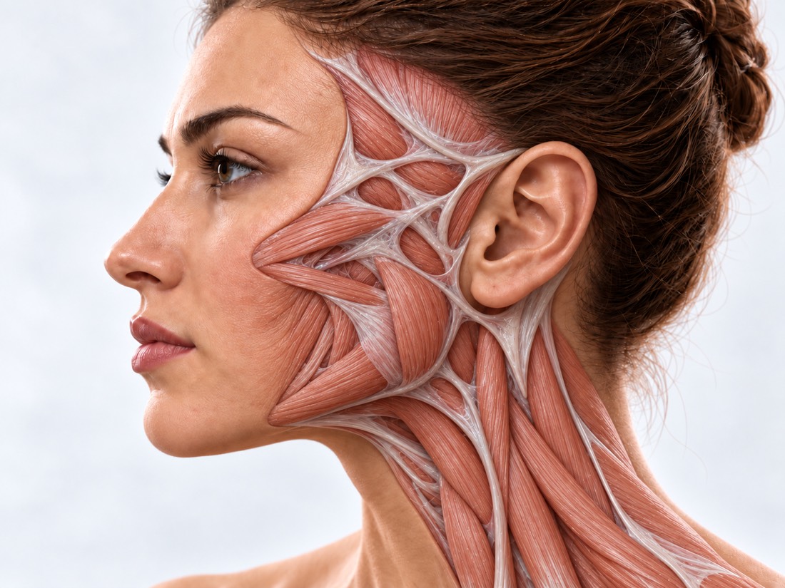 CONCEPTUAL VISUALIZATION
The bones of the face—cheekbones, orbital walls, jaws, nose, and forehead—hold the eyes, teeth, and soft tissues in place. If, after a fracture, the fragments have fused together with displacement, not only the appearance changes: the eye may become swollen, the teeth may not close properly, the face may become wider or flatter on one side.
This happens for various reasons: the operation was postponed due to injuries that are more life-threatening, the severity of the facial injury was not immediately assessed, the first operation did not completely return the debris to its place, or complications arose.
Facial surgery allows you to correct such changes both months and years after the injury. The general rule is to first return the bones to the correct position and only then work on the soft tissues and scars.
This is general information. The doctor will tell you what will suit you after the examination.
The changes depend on which bones were broken. Often a person is concerned not only with appearance, but also with how the eyes and jaws work.
One half of the face is wider, flatter or lower than the other; the cheekbone "collapsed" or protrudes.
The eyeball sits deeper or lower than on the other side. Doctors call it enophthalmos.
Fractures of the orbitDouble vision, especially when looking up or to the side, because eye movements are limited.
The teeth do not close as before the injury, it is uncomfortable to chew, the mouth does not open completely.
Asymmetry of the face and jawsThe bridge of the nose became wider, the distance between the corners of the eyes increased, there was a depression on the forehead.
Fractures of the frontal boneScarring, skin retraction, lack of volume on the cheek or near the eye.
Scars after facial injuriesDeformation is not the fault of the patient. Most often, it is related to the circumstances of the injury itself and the first treatment.
According to a meta-analysis of 78 studies, the most frequent cause of facial injuries is road accidents (55.37%), followed by seizures (17.56%) and falls (10.21%).
Half of people with facial fractures (51.04% in a meta-analysis of 26,774 patients) also had a traumatic brain injury. Then the life is saved first, and the face is operated on later.
The severity of the facial injury was not immediately assessed, and the fracture was not treated or treated inadequately.
The wreckage was not completely assembled, or complications arose. The cheekbone is the second most frequently broken facial bone after the nose; its deformations are the result of incomplete alignment, delayed surgery or an untreated fracture.
Fractures of the cheekbonesIf the injury is long-standing and the bones have already grown together, the deformity usually does not require emergency care — it is a planned situation. If the blow was recent, head trauma is first ruled out: according to the NHS, such signs require 103.
Immediately to the reception department (do not get behind the wheel) after a fresh head injury, if there was vomiting, dizziness in the head, you briefly lost consciousness or you do not remember the events surrounding the injury, the headache does not go away, you are taking blood-thinning drugs, or the injury occurred while intoxicated. On the same day: if after surgery the temperature on the face rises to 38 °C or higher or pus flows from the wound - to the surgeon on the same day. In the coming days: show a fresh fracture to the surgeon without waiting for the bones to grow: it is then easier to compare them. Planned: if the injury is long-standing, and you are concerned about asymmetry, a sore eye, diplopia, or bite, make an appointment for a consultation.
When and how the injury occurred, what operations have already been performed; medical records, old snapshots and photos of the face before the injury, if available.
The doctor evaluates the position of the eyes and orbits, the width of the cheekbones, bite, bridge of the nose, scars and skin sensitivity.
Computed tomography shows which bones have fused with displacement and to what extent. A plan cannot be made without it.
If there is double vision or an inflamed eye, the condition of the eyes is evaluated by an ophthalmologist.
For CT, the healthy half of the face is "mirrored" on the damaged one, thus obtaining a model to which the bones must be returned.
Treatment depends on which bones are affected and how long it has been. Small irregularities of the contour are sometimes leveled by simpler interventions, and a significant displacement - only by repositioning the bones. Treatment is often staged.
First, the frame of the face is turned.
Inflamed eye and diplopia.
Once the bones are in place.
Post-traumatic deformations of the face are treated at Dr. Valikhnovskyi's Institute of Surgery in the direction of "New face" - together with fresh fractures, the consequences of injuries and scars. If necessary, the condition of the eyes is monitored by an ophthalmologist.
Plastic surgeon, candidate of medical sciences, founder of Dr. Valikhnovskyi Institute of Surgery.
Short answers. What is right for you, the doctor will tell you during the consultation.
Yes. Improperly fused bones are dissected and rearranged, and the contour is leveled with grafts or implants. However, old and severe deformations are more difficult to correct than fresh ones.
Yes, repeated reconstructions are done. The doctor will evaluate the CT scan and what was done during the first surgery and make a new plan.
Not always. The sunken eye is corrected in most patients, and diplopia, according to a systematic review, has disappeared in about half. Some patients are then helped by surgery on the eye muscles or prismatic glasses.
Depends on the deformation. Bones are often restored first, and soft tissues and scars are repaired in subsequent stages. The doctor will name the number of stages after the CT scan.
A trace of the incision always remains. Access to the bones, where possible, is done in hidden places: from the side of the mouth, along the inner surface of the eyelid, behind the hairline.
It's common: Post-traumatic stress disorder, anxiety and depression are more common after facial trauma. You should tell your doctor about it.
CT images (disk or link), statements about previous operations, photo of the face before the injury. If you do not live in Kyiv, you can send them for a preliminary evaluation.
The figures on the page are from these sources.
Submit current and pre-injury facial photos and CT scans. The doctor will tell you what can be corrected and in what sequence.
Leave your contact details and briefly describe the situation. A coordinator will contact you to agree on the next step.