Pain while walking
Pain in the heel, middle, or front part of the foot, stronger under load.
 Discuss your case
Post-traumatic foot deformity — a change in the shape of the foot after a fracture or dislocation, when the bones healed incorrectly or osteoarthritis developed in the joint. It is corrected electively when pain and deformity interfere with walking and orthopedic shoes do not help.
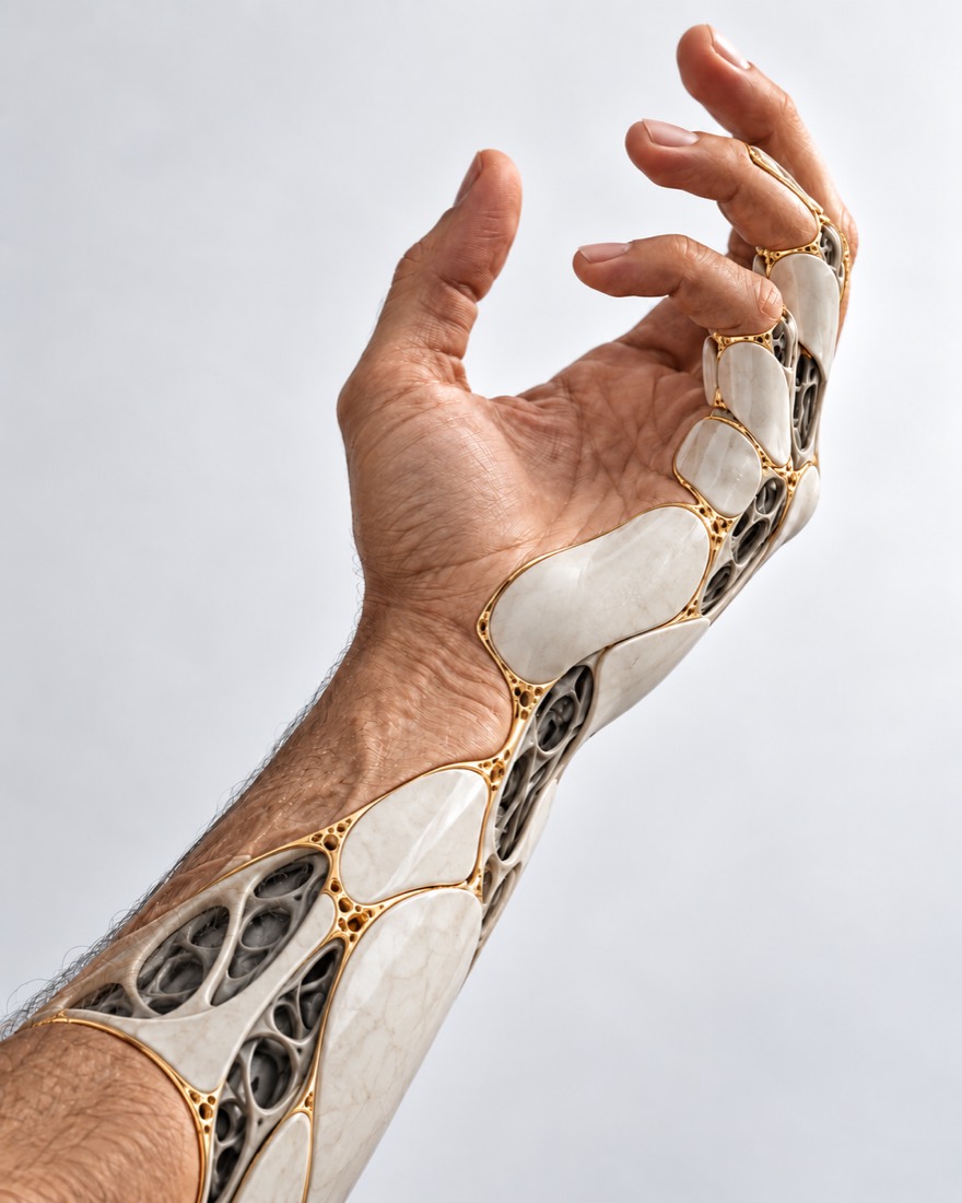 CONCEPTUAL VISUALIZATION
The foot consists of many bones and joints, which together support the body’s weight. If, after a fracture or dislocation, the bones have healed with displacement, the joint remains unstable, or arthritis has developed in it, the foot changes shape: the heel becomes lower, wider, or tilts to the side, the arch becomes flatter, and the forefoot is displaced.
These can be the consequences of fractures of the calcaneus, talus, or metatarsal bones (metatarsals account for almost 50% of foot fractures according to examinations) and injuries of the Lisfranc joint — where the middle of the foot connects with the metatarsal bones.
Such changes can be corrected even a long time after the injury. What exactly to do — whether to preserve the joint or fuse it — depends on the type of deformation and whether the cartilage has been preserved.
This is general information. The doctor will tell you what will suit you after the examination.
Changes depend on which bones and joints were damaged. Most troubling are pain while walking and uncomfortable shoes.
Pain in the heel, middle, or front part of the foot, stronger under load.
The heel is lower and wider than on the other side, tilts inward or outward; a bone may protrude on the side.
Regular shoes press or rub, making them difficult to choose.
Pain and limited movement in the foot joints — this is how post-traumatic osteoarthritis manifests.
If a metatarsal bone has fused downward, painful pressure may appear under its head.
Scars, retractions or lack of skin, tight tendons.
Soft tissue defectDeformity is not the patient's fault. It is associated with the severity of the injury, how it was recognized, and how the bones healed.
Calcaneal fractures account for up to 2% of all fractures, most often after a fall from height. After non-surgical treatment, the bone may heal with displacement, and after fractures extending into the joint, arthritis often develops in the joint under the talus.
Lisfranc joint injuries are incorrectly diagnosed in about 20% of cases upon examination. When surgeons assessed standard X-rays of 307 patients with such an injury, non-displaced injuries were recognized on average in 76.7% of cases.
In a meta-analysis of 14 studies (686 patients), osteoarthritis after injuries to the medial part of the Lisfranc joint developed in 33% during follow-up averaging about 60 months; the risk was increased by inaccurate alignment.
After talar neck fractures, according to the meta-analysis, bone necrosis developed on average in 31.2% of cases, joint osteoarthritis underneath it in 49%, and in studies with follow-up over 2 years — in 81%.
Bone defect after injuryIf the injury is old and the bones have already healed, urgent correction deformation is usually not required — it is a planned situation. Urgent signs occur after a fresh injury, cast, or surgery: according to NHS data, such signs require 103.
Immediately in the emergency department (do not drive) after a fresh injury, if the foot or ankle is at an unnatural angle, a bone is visible, there is a deep wound, or the toes are blue, pale, or numb, especially under a cast. Immediately — to the surgeon who operated or is treating the fracture, and if not possible — to the emergency room: Pain or swelling of the foot after injury or surgery is increasing, not decreasing; cast or bandage has become tight. On the same day: after a fresh injury you cannot put weight on your foot; the calf or thigh of one leg is painful and swollen (it could be a blood clot); the skin around the wound is painful, hot, and swollen; very high fever or chills; you are coughing up blood. In the coming days: if after a midfoot injury an X-ray shows nothing, but pain and swelling do not go away, — see a surgeon: a regular X-ray can miss some of these injuries, so if suspected, a CT scan is needed. Planned: if the injury is old, but the foot hurts when walking, has changed shape, or does not fit into shoes, — schedule a consultation.
When and how the injury happened, how it was treated, what surgeries were performed; discharge summaries and old images.
The doctor assesses the shape of the foot, gait, joint movements, skin and scars, circulation, and sensitivity.
X-ray shows the position of the bones, CT shows exactly how they have fused and whether the joint surfaces are damaged. If Lisfranc injury is suspected, but the X-ray shows nothing, a CT scan is performed.
It determines whether the joint can be preserved or if it is better to fuse it.
With CT, the angles and cut lines can be pre-modeled in a special program.
Treatment depends on which bones are affected, whether the cartilage is preserved, and how much the deformity interferes with walking. The goal is to reduce pain and restore support to the foot.
The first step if the deformity is moderate.
If the cartilage is still intact.
If the joint is destroyed by osteoarthritis.
Post-traumatic foot deformities at the Valikhnovskyi Institute of Surgery are treated under the 'Hand and Foot Restoration' program. Surgery is planned when pain or deformity interferes with walking and orthopedic shoes do not help; the plan is made based on examination and CT scan.
Plastic surgeon, candidate of medical sciences, founder of Dr. Valikhnovskyi Institute of Surgery.
Short answers. What is right for you, the doctor will tell you during the consultation.
Yes. Bones that have healed in displacement are cut and repositioned, and if the joint is destroyed by arthritis, it is fixed in the correct position.
If the pain is moderate, orthopedic shoes and insoles help. Surgery is discussed when they no longer help and the pain and deformity interfere with walking.
It is the fusion of a joint: the bones are joined together in the correct position. This eliminates arthritis in the joint, but movement in it is lost.
Damage in the middle of the foot, in particular the Lisfranc joint, is not always visible on a regular X-ray, especially without displacement. Therefore, if suspected, a CT scan is done.
For large defects, individual titanium 3D implants are used. Whether this is suitable for you will be told by the doctor after a CT scan.
After foot surgery, for several weeks one walks in special shoes with limited weight-bearing. The exact timing depends on the surgery.
X-ray and CT scans (disc or link), records of fracture treatment and surgery, photos of the feet. If you do not live in Kyiv, they can be sent for preliminary evaluation.
The figures on the page are from these sources.
Send photos of the feet, X-ray, and CT scan. The doctor will say what can be corrected and whether it is possible to preserve the joint.
Leave your contact details and briefly describe the situation. A coordinator will contact you to agree on the next step.