CONDITION / TUMOURS

Sealing under the skin.

A subcutaneous lump — a nodule or "ball" in the skin, adipose tissue, or muscle, the nature of which has not yet been determined. Most often it is something benign, but some signs are a reason to be examined without delay.
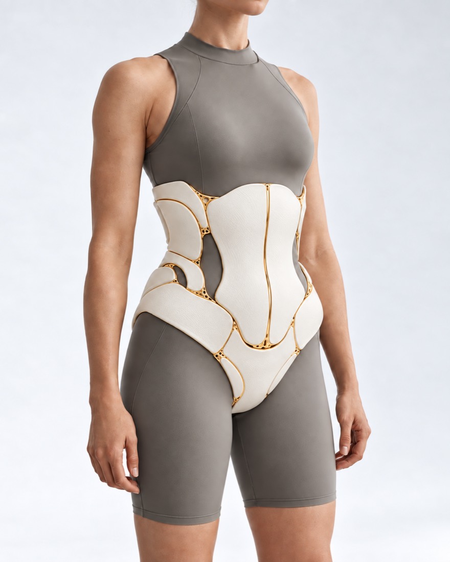
Conceptual visualization: soft tissues of the trunk where the doctor searches for a lumpCONCEPTUAL VISUALIZATION
What it is
A nodule under the skin or in the muscle, the nature of which has not yet been determined.
How often is it cancer
Among 2,399 people referred for ultrasound by family doctors due to a lump, malignant tumors were found in 0.7%.
Alarming signs
Larger than 5 cm, growing, located deep under the fascia, reappeared at the site of the removed one.
Main rule
If there is suspicion of sarcoma, first MRI and biopsy, and then surgery in a center that treats sarcomas.

What is
Lump under the skin.

A lump under the skin is the general term for any nodule you feel under the skin or in the muscle. It is not a diagnosis: it may appear as a lipoma (fat tumor), cyst, fibroma, inflammation, or, more rarely, a malignant tumor.

Most lumps are benign. Malignant soft tissue tumors — sarcomas — are rare: in Europe, their incidence is 4–8 cases per 100,000 people. But a sarcoma can easily be mistaken for a benign nodule, and then it is removed like a regular lipoma — without a margin of healthy tissue.

Therefore, the order of actions with a lump is important: first, find out what it is, and only then decide whether to remove it and how. For most people, an examination and ultrasound are sufficient; if there are worrying signs, MRI and biopsy are needed.

This page is about a lump whose nature has not yet been determined. If the diagnosis is already known, see the pages about lipomas or soft tissue sarcomas.

This is general information. The doctor will tell you what will suit you after the examination.

How does it look
Lump under the skin.

The appearance of a lump gives a clue as to what it might be, but it is impossible to determine its exact nature by touch.

02

Smooth under the skin

A smooth nodule just under the skin may be a cyst.

03

It hurts, red, hot

A swollen painful lump, with the skin over it red or hot — this is what an abscess, a purulent inflammation, looks like.

04

Solid and immovable

A hard lump that is difficult to move under the skin should be shown to a doctor.

05

It grows

A lump is increasing without an obvious reason — a reason to do an ultrasound within 2 weeks.

06

Deep and large

Lies deep, under the muscle layer (fascia), or is more than 5 cm. A sarcoma usually does not hurt at first and grows.

Soft tissue sarcoma

What can it do?
to be

There are many causes. Mostly these are benign formations, but excluding a malignant tumor without examination is impossible.

Cysts and fibromas

Skin cysts, ganglions near joints, fibromas — are also benign formations.

Inflammation

Abscess — a cavity with pus after an infection, usually painful and red.

Sarcoma

Malignant tumor of muscles, fat, or connective tissue. Doctors distinguish about 80 types of it.

Soft tissue sarcoma

What raises suspicion

In a series of 280 patients referred with suspected malignant tumor, deep location, size over 5 cm, age over 50 years, and location closer to the trunk on the arm or leg together increased the probability of sarcoma up to 50%.

When to seek medical advice?

A subcutaneous lump usually does not require emergency care. The important thing is different: not to delay examination if there are alarming signs, and to notice the signs of severe infection in time.

103 immediately if:

  • person breathes very frequently;
  • confusion, unclear speech, the person speaks nonsense;
  • skin is blue, pale, or blotchy;
  • very high or very low body temperature;
  • a rash that does not blanch when pressed;
  • a child under 5 breathes very fast or with difficulty: grunting, belly pulls in under the ribs;
  • a child under 5 has blue, gray, pale, or mottled skin, lips, or tongue;
  • a child under 5 has a rash that does not blanch when pressed;
  • a child under 3 months has a temperature of 38 °C and above, at 3–6 months — 39 °C and above;
  • a child under 5 has a temperature below 36 °C, is cold to the touch or shivering;
  • a child under 5 cries weakly, piercingly, or continuously;
  • a child under 5 responds poorly, does not eat, is difficult to wake, or is sleepier than usual;
  • a child under 5 has not urinated for 12 hours.

Do not delay a visit to the doctor:

On the same day: a lump is painful, red, or hot; redness or swelling spreads from it; you feel chills or feverish. Within 2 weeks — see a doctor: A lump grows without a clear reason (in that case, an ultrasound is needed within the same 2 weeks), larger than 5 cm, lies deep, is hard and immobile, or has reappeared at the site of a removed one. A child or young person up to 24 years old with a growing lump should have an ultrasound within 48 hours. See a doctor as soon as possible: any lump that does not resolve for more than 2 weeks. Planned: a soft, long-standing lump that does not change but is bothersome.

As it turns out,
what is it

  1. Review

    The doctor asks when the lump appeared and whether it is growing, measures it, assesses how deep it lies and whether it moves. But it is difficult to determine the nature of a node by touch: in a study of 823 superficial formations, the accuracy of examination in determining the type of formation was 25.6%, ultrasound — 81.2%.

  2. ultrasound

    First examination. In a study of 2,399 people referred from primary care, ultrasound showed a benign pattern in 95%, and 91% required no further action. For deep lesions, ultrasound detected 97% of malignant ones but also considered some benign lesions suspicious.

  3. MRI with contrast

    If there are alarming signs or a suspicious ultrasound pattern, an MRI with contrast is performed. It shows where the tumor is and how it is separated from muscles and other tissues.

  4. Biopsy with a thick needle

    Small pieces of tissue are taken with a needle for examination under a microscope. Guidelines recommend biopsy for all deep lesions and superficial ones larger than 3 cm; there are separate rules for fatty tumors.

  5. Sarcoma center

    If ultrasound indicates sarcoma or the result is unclear, and concern remains, the person is referred to a center that treats sarcomas. The diagnosis there is confirmed with MRI and biopsy.

What are they doing?
with a lump.

Treatment depends on what the examination showed. Here are three main approaches.

01

Benign

Lipoma, cyst, fibroma.

Follow-up
If the diagnosis is clear and the nodule does not interfere, removal is not necessary.
Removal
A lipoma or cyst is removed if it hurts, gets in the way, grows, or there is doubt about its nature.
02

Inflammation

Abscess.

Incision
Pus is usually released through a small incision; small abscesses are sometimes treated with antibiotics only.
03

Suspicion of sarcoma

Do not remove "just like that".

Why not to enucleate
In a meta-analysis of 16,946 patients with sarcoma, more than a third were initially operated on as a benign nodule. After such unplanned removal, the tumor more often returned in the same place.
What's next
After unplanned removal, a repeat, wider surgery at the tumor site is usually required. Unplanned removal was not associated with worse overall five-year survival in a meta-analysis, but residual tumor in the wound was associated with more frequent local recurrence.
Where to treat
Guidelines recommend that if sarcoma is suspected, the person should be referred to a specialized center where they are managed by a team of a surgeon, oncologist, and radiation therapist.
How we treat
After confirming the diagnosis, the main treatment is the removal of the tumor by an experienced surgeon, with radiation therapy before or after surgery as indicated.

How
we treat it.

A lump under the skin at the Valikhnovskyi Institute of Surgery is examined and treated in the 'Soft Tissue Oncoplastic Surgery' department: the clinic is responsible for examination, biopsy, removal, and tissue reconstruction. If sarcoma is confirmed, treatment is planned together with an oncologist and a radiation therapist.

CLINIC SERVICE Oncosurgery of soft tissues Sarcomas and other soft tissue tumors — with preservation of the limb and restoration of tissues in the same operation. View service

What we do for this condition

  • MRI and biopsy with a thick needle — with histology before surgery; intervention is planned only after diagnosis.
  • Consilium — the surgeon, oncologist, and radiation therapist decide whether preoperative treatment is necessary.
  • 3D modeling — if needed, MRI and CT data are combined into a three-dimensional model to visualize the tumor in relation to blood vessels, nerves, and bones.
  • Single block deletion — the tumor is removed along with a layer of healthy tissue without opening it; a pathologist checks the margins.
  • Closure of the defect with a flap — if the wound cannot be simply sutured, — often during the same surgery.

Rostislav Valikhnovskyi

Plastic surgeon, candidate of medical sciences, founder of Dr. Valikhnovskyi Institute of Surgery.

How treatment works with us

  1. Materials onlineSend a photo of the lump, ultrasound or MRI reports, and biopsy results, if available.
  2. ConsultationExamination, assessment of tests, and a decision on whether MRI and biopsy are needed.
  3. DiagnosisMRI and core needle biopsy; histology shows what kind of formation it is.
  4. PlanObservation, simple removal, or, in case of sarcoma, a council and surgery plan.
  5. Surgery and follow-upRemoval with tissue examination, followed by scheduled check-ups.

Common
questions

Short answers. What is right for you, the doctor will tell you during the consultation.

The lump doesn’t hurt — does that mean it’s not dangerous?

No. A sarcoma usually does not hurt at first. In a study of 135 urgent referrals, pain was the least useful sign: 27% of people with a malignant tumor had it.

Can you just cut out the lump without examining it?

It’s not worth it if there are warning signs. If it’s a sarcoma, removing it without a margin of healthy tissue increases the risk of recurrence and may require a repeat, more extensive surgery.

How many lumps turn out to be cancer?

Few. Among 2,399 people referred for ultrasound by family doctors, 0.7% had malignant tumors. Among 135 people urgently referred within a year to one UK center due to warning signs, 33% did.

Is an ultrasound enough?

It often happens: if the lesion is clearly benign, nothing else is needed. If the ultrasound shows a suspicious or unclear picture, MRI and biopsy are performed.

Why a needle biopsy and not an incision?

In a meta-analysis of 17 studies (2,680 patients), core needle biopsy identified 97% of malignant and 99% of benign lesions and caused fewer complications than incision biopsy. The authors note that the quality of the studies is low.

A lump in a child – what to do?

See a doctor. If the lump grows without a clear reason, an ultrasound should be done within 48 hours.

A lump appeared again after removal. What to do?

See a doctor within 2 weeks: recurrence at the site of removal is one of the signs for which waiting is not advisable. The doctor will decide whether ultrasound, MRI, and biopsy are needed.

Sources

19 sources · guidelines, meta-analyses, studies, NICE and NHS
showCollapse
  1. UK guidelines for the management of soft tissue sarcomasBritish Journal of Cancer, 2025doi:10.1038/s41416-024-02674-y ↗
  2. ASO Practice Guidelines Series: Soft Tissue Sarcoma of the Extremities and Superficial TrunkAnnals of Surgical Oncology, 2025doi:10.1245/s10434-025-18223-7 ↗
  3. Diagnosis strategy of adipocytic soft-tissue tumors in adults: a consensus from European expertsEuropean Journal of Surgical Oncology, 2022doi:10.1016/j.ejso.2021.10.009 ↗
  4. Soft Tissue Masses: Evaluation and TreatmentAmerican Family Physician, 2022pubmed.ncbi.nlm.nih.gov/35704829 ↗
  5. Evaluating the British sarcoma referral formAnnals of the Royal College of Surgeons of England, 2015doi:10.1308/rcsann.2015.0002 ↗
  6. Soft-tissue masses of the locomotor system. A guide to the clinical diagnosis of malignancyActa Orthopaedica Scandinavica, 1986doi:10.3109/17453678608994379 ↗
  7. An assessment of Ultrasound screening for soft tissue lumps referred from primary careClinical Radiology, 2018doi:10.1016/j.crad.2018.07.102 ↗
  8. Accuracy of ultrasound in the characterization of superficial soft tissue tumors: a prospective studySkeletal Radiology, 2020doi:10.1007/s00256-019-03365-z ↗
  9. Accuracy of ultrasound in the characterisation of deep soft tissue masses: a prospective studyEuropean Radiology, 2020doi:10.1007/s00330-020-07002-5 ↗
  10. Core needle biopsy versus incisional biopsy for differentiation of soft-tissue sarcomas: A systematic review and meta-analysisCancer, 2020doi:10.1002/cncr.32735 ↗
  11. Is Unplanned Excision of Soft Tissue Sarcomas Associated with Worse Oncological Outcomes? A Systematic Review and Meta-AnalysisCancers, 2024doi:10.3390/cancers16020443 ↗
  12. Impact of NICE guidelines on the survival of patients with soft-tissue sarcomasBone & Joint Journal, 2021doi:10.1302/0301-620X.103B3.BJJ-2020-0743.R1 ↗
  13. Imaging recommendations for soft-tissue sarcomas: model guidelines from diagnosis to post-treatment follow-upClinical Radiology, 2024doi:10.1016/j.crad.2024.08.023 ↗
  14. Suspected cancer: recognition and referral (NG12) — sarcomasNICE, 2026nice.org.uk/guidance/ng12/chapter/Recommendations-organised-by-site-of-cancer ↗
  15. Suspected cancer: recognition and referral (NG12) — terms usedNICE, 2026nice.org.uk/guidance/ng12/chapter/Terms-used-in-this-guideline ↗
  16. LumpsNHS, 2024nhs.uk/symptoms/lumps ↗
  17. Symptoms of soft tissue sarcomaNHS, 2024nhs.uk/conditions/soft-tissue-sarcoma/symptoms ↗
  18. Skin abscessNHS, 2026nhs.uk/conditions/skin-abscess ↗
  19. SepsisNHS, 2026nhs.uk/conditions/sepsis ↗

The figures on the page are from these sources.

VALIKHNOVSKI SURGERY PARTNERS

Noticed a lump,
that is growing?

Send a photo and ultrasound or MRI reports, if available. The doctor will say which tests are needed and whether removal is advised.

Call Send an enquiry
ONLINE ENQUIRY

Tell us how we can help.

Leave your contact details and briefly describe the situation. A coordinator will contact you to agree on the next step.

Preferred contact method
