CONDITION / TUMOURS

Soft tissue sarcoma.

Soft tissue sarcoma is a rare malignant tumor of muscles, fat, or connective tissue. Most often it appears as a growing lump. The main treatment is surgery, often together with radiation therapy; the plan is made by a team of specialists.
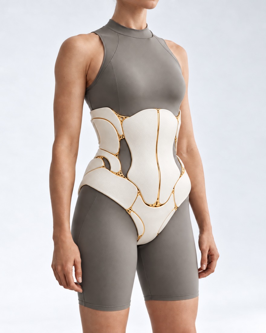
Conceptual visualization: body soft tissues and their recovery after tumor removalCONCEPTUAL VISUALIZATION
What it is
A rare malignant tumor of muscles, fat, or other connective tissue.
How common
About 1% of all malignant tumors in adults; in Europe — 4–8 cases per 100,000.
The main feature
A growing lump — usually deep under the skin, firm, initially painless.
Who treats it
Team: surgeon, oncologist, radiation therapist; surgery and tissue reconstruction are performed by the surgeon.

What is
soft tissue sarcoma.

Soft tissue sarcoma is a rare cancer that begins in muscles, fat tissue, or other connective tissues. It can occur almost anywhere in the body. There are over 100 types, and it most often grows on the arms and legs, trunk, and in the retroperitoneal space.

Lumps and swelling under the skin occur often and have many causes, so a lump by itself does not necessarily mean sarcoma. But it is worth showing it to a doctor: if it is cancer, early detection can make treatment easier.

Because sarcomas are rare and very different, guidelines recommend treating them in centers where there is a team with experience specifically in these tumors — specialists from various fields.

If there is no diagnosis yet and you have only noticed a lump, see the page 'Lump under the skin.' About recurrence of the tumor after treatment — see the page 'Sarcoma recurrence.'

This is general information. The doctor will tell you what will suit you after the examination and examinations.

How
soft tissue sarcoma presents.

The most common sign is a lump on the body. Other signs depend on exactly where the tumor is growing.

02

Deep under the skin

Usually lies deep, so it can be felt before it can be seen.

03

Hard and immobile

Feels firm and does not move easily under the skin.

04

Does not hurt initially

Usually does not hurt; as it grows, it may start to hurt.

05

Signs near organs

Abdominal pain and constipation — if the tumor is near the abdomen; a cough that does not go away — if it is near the lungs.

Who has
higher risk.

Why sarcoma occurs is not always known. But there are conditions under which it happens more often.

age

The risk is higher after 65 years of age.

Previous radiation exposure

Radiation therapy that was once carried out for another disease.

Retinoblastoma

A rare eye cancer in childhood.

Untreated HIV

Can cause a special type of tumor — Kaposi's sarcoma.

When to seek medical advice?

A growing lump is a reason to see a doctor without delay, but it is not necessary to call an ambulance because of it. 103 may be needed due to complications after surgery.

103 immediately if:

  • bleeding from the wound does not stop, even if it is pressed;
  • blood gushes, bright red, it is difficult to stop;
  • suddenly having difficulty breathing;
  • chest pain or pain in the upper back;
  • the heart beats very often;
  • the person fainted.

To the doctor:

Immediately - to the surgeon who operated, and if it does not work - to the waiting room: After surgery, pain or swelling in the surgical area increases; the calf or leg hurts and swells. On the same day: The wound has become red, pus is coming out; you feel unwell or have a fever; a cough with blood appears. Within 48 hours: If a child or teenager has a lump that is growing without a clear reason — an ultrasound is needed very urgently. Without delay, by appointment: If an adult has a lump that is growing without a clear reason — an ultrasound should be done within 2 weeks; if the ultrasound shows suspicion of sarcoma or the result is unclear and doubts remain, — go to the team that treats sarcomas: MRI and biopsy are done where the surgery will take place. Planned: Any new lump or swelling on the body should be shown to a doctor.

How to make a diagnosis
Before surgery.

  1. Examination and Ultrasound

    The doctor examines the lump and, if necessary, orders blood tests and an ultrasound.

  2. MRI or CT

    They show the size of the tumor and how it lies in relation to the muscles, blood vessels, nerves, and bones. They are used as a basis when planning biopsy and surgery.

  3. Biopsy with a thick needle

    Diagnosis is made only through tissue examination. In a meta-analysis of 17 studies (2,680 patients), such a biopsy detected malignant tumors in 97% of cases and caused fewer complications than an incisional biopsy. It is recommended to carefully plan the biopsy before surgery — by the team that will treat the sarcoma.

  4. Type and spread

    Additional CT scans, PET-CT if needed, and genetic analyses of the tumor show what type of sarcoma it is and whether it has spread. The treatment plan depends on this.

How we treat
soft tissue sarcoma.

The main treatment is surgery, aimed at removing the tumor with clear margins while preserving limb function. Radiation and medications are added depending on the type and risk of the tumor. The plan is made by a team of specialists.

01

Where to receive treatment

At a center experienced in treating sarcomas.

Team of specialists
The British guideline recommends referring every patient suspected of sarcoma to a specialized team. According to the European ESTRO guideline, discussion in an expert center remains mandatory to choose the treatment.
What this gives
In the French registry of 35,784 patients, surgery in a reference center was associated with a lower risk of tumor recurrence and death.
02

Surgery

Main treatment.

Single block deletion
The tumor is removed together with a layer of healthy tissue without opening it. The goal is clean margins, without tumor cells at the edge of the incision.
Why not 'shelled out'
When a tumor is removed without examination and tissue margins, in most cases tumor cells remain in the wound. In a meta-analysis, residual tumor increased the likelihood of local recurrence (odds ratio 3.56). Therefore, after such surgery, a wider repeat surgery is often performed.
Limb preservation
In a randomized study of 43 patients, limb-sparing surgery with radiation resulted in a 5-year survival of 83% compared to 88% after amputation — the difference was not significant. Local recurrences after limb-sparing were 4, after amputation — none; the only factor associated with recurrence was margin status.
03

Radiation and medication

They are prescribed by an oncologist and a radiation therapist.

Why radiation
It is often added to reduce the risk of the tumor returning in the same place. The European ESTRO 2026 guideline prefers preoperative radiation.
Before or after surgery
In a randomized study of 190 patients, after preoperative radiation, the wound was more often complicated — in 35% versus 17%. Therefore, the scheme is chosen based on the size and location of the tumor.
Chemotherapy
Not everyone needs it: it is considered for types of sarcomas sensitive to drugs, and in certain high-risk patients. The decision is made by the oncologist.
04

Tissue reconstruction

Often — in the same operation.

Patches
If the wound cannot be simply stitched, it is closed using the patient's own tissue on vessels. In a systematic review of 90 studies, the limb was preserved in more than 90% of cases with all types of reconstruction.
Joint planning
According to the same review, joint planning between the oncologic surgeon and the plastic surgeon is associated with fewer wound complications.
Immediately or later
In one retrospective study of 70 patients, delayed reconstruction (after more than 3 weeks) more often led to infection and wound dehiscence compared to immediate reconstruction.
05

After treatment

Supervision
Regular check-ups are needed to notice tumor recurrence, metastases, and late treatment effects in time.
Examination
The surgical area is monitored using MRI, the lungs — by chest CT. The intervals depend on the degree of malignancy and the type of tumor.

How
we treat it.

Soft tissue sarcoma at the Dr. Valikhnovskyi Institute of Surgery is treated in the direction of "Soft Tissue Onco-Surgery." The clinic is responsible for the surgical part — tumor removal and restoration of tissues, blood vessels, and nerves. Chemotherapy and radiation therapy are prescribed and managed by an oncologist and radiation therapist; the treatment plan is developed together, at a consultation.

CLINIC SERVICE Oncosurgery of soft tissues Sarcomas and other soft tissue tumors — with preservation of the limb and restoration of tissues in the same operation. View service

What we do for this condition

  • MRI and biopsy with a thick needle — first the diagnosis is made by histology, only then — the operation planning.
  • Consilium — the surgeon, oncologist, and radiation therapist decide whether preoperative treatment is necessary.
  • 3D modeling — if necessary, MRI and CT data are combined into a three-dimensional model to visualize the vessels, nerves, and bones near the tumor.
  • En bloc removal — the tumor is removed along with the pseudocapsule and a layer of healthy tissue; the edges are evaluated by a pathologist.
  • Limb preservation — the goal is to remove the tumor completely while preserving the arm or leg and its function.
  • Vascular prosthetics and nerve repair — the affected artery or vein is replaced with a graft or the patient’s own vein; the nerve, if possible, is reconstructed.
  • Flaps in the same surgery — large defects are closed with local flaps on a vascular pedicle or with microsurgical anastomoses.
  • Rehabilitation — after muscle removal and nerve repair — exercises to restore strength and movement.

Rostislav Valikhnovskyi

Plastic surgeon, candidate of medical sciences, founder of Dr. Valikhnovskyi Institute of Surgery.

How treatment works with us

  1. Materials onlineSend MRI, CT, biopsy report, and discharge summaries if available.
  2. ConsultationExamination and assessment of tests; if needed — MRI and core needle biopsy.
  3. Consilium and planTogether with an oncologist and radiation therapist: determine if preoperative treatment is needed, the extent of removal, and how to close the wound.
  4. SurgeryEn bloc tumor removal and tissue reconstruction — more often in a single operation.
  5. Recovery and supervisionHealing, rehabilitation, then — regular chest MRI and CT scans as scheduled by the oncologist.

Common
questions

Short answers. What is right for you, the doctor will tell you during the consultation.

Is every lump under the skin a sarcoma?

No. Lumps often occur and have many causes, while sarcoma is a rare cancer. But a lump that is growing should be shown to a doctor as soon as possible.

Can a lump just be cut out?

It is not recommended without examination. If it is a sarcoma, after such removal, tumor cells usually remain in the wound, and a repeated, wider surgery is needed. First — MRI and biopsy.

Why a biopsy if the tumor will be removed anyway?

The type of sarcoma determines the plan: whether radiation or medication is needed before surgery and exactly how to operate. A biopsy is recommended to be carefully planned before surgery — with the team that will treat the sarcoma.

Is it possible to save the arm or leg?

The goal of surgery is to remove the tumor with clear margins and preserve the function of the limb. In a systematic review of 90 studies among patients who had tissue reconstruction with a flap after tumor removal from bones or soft tissues, the limb could be preserved in more than 90% of cases. Whether this is possible for you specifically depends on the size and location of the tumor.

Why is it important to be treated in a specialized center?

In a French registry of over 35,000 patients, surgery in a reference center was associated with a lower risk of recurrence and death. Therefore, guidelines recommend that treatment be planned by a team with experience in treating sarcomas.

Is chemotherapy mandatory?

No. The main treatment is surgery, often with radiation. Chemotherapy is considered for certain types of sarcomas and high risk; the decision is made by the oncologist.

Is reconstruction done immediately?

If the wound cannot be simply stitched, it is often covered with a flap in the same surgery. In one study, delayed reconstruction more often led to infection and wound dehiscence.

How often to be examined after treatment?

Regularly: MRI of the surgical area and CT of the chest. Intervals depend on the malignancy grade and type of tumor; the schedule is determined by the oncologist.

Sources

21 sources · British, European, and American guidelines, meta-analyses, randomized studies, NHS and NICE
showCollapse
  1. UK guidelines for the management of soft tissue sarcomasBritish Journal of Cancer, 2025doi:10.1038/s41416-024-02674-y ↗
  2. ESTRO clinical practice guideline on radiotherapy for adult soft tissue sarcoma of the extremities and trunk wall – Endorsed by ASTRORadiotherapy and Oncology, 2026doi:10.1016/j.radonc.2026.111535 ↗
  3. ASO Practice Guidelines Series: Soft Tissue Sarcoma of the Extremities and Superficial TrunkAnnals of Surgical Oncology, 2025doi:10.1245/s10434-025-18223-7 ↗
  4. Soft-tissue sarcoma in adults: An update on the current state of histiotype-specific management in an era of personalized medicineCA: A Cancer Journal for Clinicians, 2020doi:10.3322/caac.21605 ↗
  5. Imaging recommendations for soft-tissue sarcomas: model guidelines from diagnosis to post-treatment follow-upClinical Radiology, 2024doi:10.1016/j.crad.2024.08.023 ↗
  6. Diagnosis and treatment of primary soft tissue sarcomas: comprehensive reviewBJS Open, 2026doi:10.1093/bjsopen/zraf177 ↗
  7. Surgery in reference centers improves survival of sarcoma patients: a nationwide studyAnnals of Oncology, 2019doi:10.1093/annonc/mdz124 ↗
  8. Improved nationwide survival of sarcoma patients with a network of reference centersAnnals of Oncology, 2024doi:10.1016/j.annonc.2024.01.001 ↗
  9. Core needle biopsy versus incisional biopsy for differentiation of soft-tissue sarcomas: A systematic review and meta-analysisCancer, 2020doi:10.1002/cncr.32735 ↗
  10. Re-excision after unplanned excision of soft tissue sarcoma: A systematic review and metanalysis. The rationale of systematic re-excisionJournal of Orthopaedics, 2021doi:10.1016/j.jor.2021.05.022 ↗
  11. The treatment of soft-tissue sarcomas of the extremities: prospective randomized evaluations of (1) limb-sparing surgery plus radiation therapy compared with amputation and (2) the role of adjuvant chemotherapyAnnals of Surgery, 1982doi:10.1097/00000658-198209000-00009 ↗
  12. Preoperative versus postoperative radiotherapy in soft-tissue sarcoma of the limbs: a randomised trialThe Lancet, 2002doi:10.1016/S0140-6736(02)09292-9 ↗
  13. Soft tissue reconstruction techniques after musculoskeletal tumor resection: A systematic review of gastrocnemius flap, free flap, local flap, and orthoplastic reconstructionEuropean Journal of Surgical Oncology, 2026doi:10.1016/j.ejso.2026.112077 ↗
  14. Soft tissue tumor imaging in adults: European Society of Musculoskeletal Radiology-Guidelines 2024: imaging immediately after neoadjuvant therapy in soft tissue sarcoma, soft tissue tumor surveillance, and the role of interventional radiologyEuropean Radiology, 2025doi:10.1007/s00330-024-11242-0 ↗
  15. Immediate versus Delayed Sarcoma Reconstruction: Impact on OutcomesSarcoma, 2016doi:10.1155/2016/7972318 ↗
  16. Symptoms of soft tissue sarcomaNHS, 2024nhs.uk/conditions/soft-tissue-sarcoma/symptoms ↗
  17. Causes of soft tissue sarcomaNHS, 2024nhs.uk/conditions/soft-tissue-sarcoma/causes ↗
  18. Suspected cancer: recognition and referral (NG12). SarcomasNICE, 2026nice.org.uk/guidance/ng12/chapter/Recommendations-organised-by-site-of-cancer ↗
  19. Cuts and grazesNHS, 2026nhs.uk/conditions/cuts-and-grazes ↗
  20. Pulmonary embolismNHS, 2026nhs.uk/conditions/pulmonary-embolism ↗
  21. DVT (deep vein thrombosis)NHS, 2026nhs.uk/conditions/deep-vein-thrombosis-dvt ↗

The figures on the page are from these sources.

VALIKHNOVSKI SURGERY PARTNERS

Noticed a lump,
that is growing?

Send an MRI, biopsy conclusion, or discharge summary if available. The doctor will say which examinations are missing and how the tumor can be removed while preserving function.

Call Send an enquiry
ONLINE ENQUIRY

Tell us how we can help.

Leave your contact details and briefly describe the situation. A coordinator will contact you to agree on the next step.

Preferred contact method
