CONDITION / FACE

Vascular malformations of the face and neck.

Vascular malformations of the face and neck are defects in the structure of blood or lymphatic vessels with which a person is born. Lymphangioma belongs to them. This is not a hemangioma: the treatment is different, and it is often conducted by several specialists together.

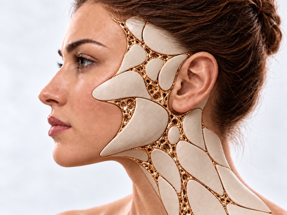
Conceptual visualization: soft tissues of the face and neckCONCEPTUAL VISUALIZATION
What it is
Defect in the structure of blood vessels: is present from birth, although it may become noticeable later.
Types
Capillary ("wine stain"), venous, lymphatic ("lymphangiomas") and arteriovenous; are mixed.
Where
Most often — on the head and neck; about 75% of lymphatic malformations are located there.
Who treats it
Team: surgeon, interventional radiologist, ENT doctor and dermatologist if necessary.

What is
vascular malformation.

Vascular malformation is a defect in the structure of blood or lymphatic vessels that occurs before birth. It is present from birth, although it may become noticeable later. Most often, such defects occur on the head and neck.

According to the classification of ISSVA - the International Society for the Study of Vascular Anomalies - malformations are a separate group, and hemangioma belongs to vascular tumors. Mislabeling and misdiagnosis are common, and treatment depends on an accurate diagnosis.

According to the type of vessels, there are capillary, venous, lymphatic and arteriovenous malformations; if several types of vessels are affected - mixed. Lymphangioma is the old name of a lymphatic malformation. About 90% of lymphatic malformations are detected before age 2, and about 75% of them are located in the head and neck. They are benign, but rarely disappear on their own.

Treatment of malformations is the work of a team: a surgeon, an interventional radiologist, and, if necessary, an ENT doctor and a dermatologist. It is not always possible to completely cure the malformation; then the goal is to reduce symptoms and improve quality of life.

This is general information. What will suit you or your child, the doctor will tell after the examination.

How they manifest
malformations

Appearance and symptoms depend on which vessels are affected, where and how deep.

01

Wine stain

A capillary malformation is a red, purple, or dark spot, most often on the face and neck, usually on one side. Is from birth; can darken and become bumpy without treatment.

02

Venous malformation

Swelling from varicose veins. It can hurt and swell, blood clots can form in it. On the lips, mouth or tongue, it is sometimes combined with changes in the respiratory tract.

03

Lymphangioma

Lymphatic malformation is a soft formation with cyst cavities: large, small or mixed. It can increase rapidly after an infection, injury, or hemorrhage.

04

Arteriovenous malformation

Arteries connect directly to veins, blood flows through them quickly. Typical places on the head are the face and oral cavity.

05

Effect on functions

Large malformations of the face and neck change the appearance and can interfere with breathing, swallowing and speaking.

Why do they arise?
and what makes them stronger.

The malformation is laid before birth. But some circumstances can dramatically increase it or cause it to return.

Vascular malformation

Blood vessels are formed incorrectly even before birth. Therefore, the malformation is neither a tumor nor a hemangioma.

Facial hemangiomas

Infection, trauma, hemorrhage

A lymphatic malformation can quickly enlarge and press on the airways, making it difficult to swallow and speak.

Hormonal changes

The growth of arteriovenous malformation during pregnancy is described.

Incomplete deletion

An arteriovenous malformation often returns if it is not completely removed or occluded.

Combination with syndromes

A wine stain is occasionally a sign of a syndrome, such as Sturge-Weber; then additional examinations are required.

When to seek medical advice?

Most malformations are long-standing, planned conditions, they usually do not require emergency care. But a malformation in the neck, mouth, or tongue can suddenly enlarge after infection, trauma, or hemorrhage and compress the airway.

103 immediately if:

  • it became difficult to breathe;
  • cannot swallow or saliva flows;
  • a high-pitched whistling sound (stridor) is heard during inhalation;
  • swelling on the neck, mouth or tongue grows rapidly;
  • bleeding that cannot be stopped;
  • the blood gushes out — bright red, hard to stop.

Do not delay a visit to the doctor:

On the same day: the malformation has suddenly become larger, painful, red or hot, or has a high temperature - this could be an infection or haemorrhage. Planned: a spot or swelling that has been there for a long time and does not change quickly; a wine stain in a child — the laser gives a greater effect in small children; a malformation near the eye, nose, or mouth that grows or hurts.

How to determine
type of malformation.

  1. Discussion and review

    When it was noticed whether the size changes, whether there were infections, bleeding, sudden increases.

  2. MRI

    Basic examination: shows the type of malformation, its boundaries and the relationship with neighboring tissues.

  3. Ultrasound and angiography — if necessary

    Ultrasound complements the examination; in case of arteriovenous malformation, vessels are examined by angiography before treatment.

  4. Examination of the respiratory tract

    In case of malformations of the lips, mouth, tongue or neck, the ENT doctor evaluates the respiratory tract, even if there are no complaints about breathing.

How we treat
malformations

Treatment depends on the type, size and location of the malformation. It is selected individually, often carried out in stages and combines several methods.

01

Lymphatic (lymphangiomas)

Sclerotherapy, surgery or their combination.

Sclerotherapy
Through a needle, a drug is injected into the cysts, which glues their walls together; it is performed by interventional radiologists. According to a meta-analysis of 25 studies (726 patients), complete cure was achieved in 50.5%: with large cysts — in 53.1%, with small cysts — in 35.1%, and with mixed cysts — in 31.1%. Persistent complications — 1.2%.
Surgery
With large cysts in a series of 63 children, one operation completely removed the malformation in 90.5%. Complications: fluid or blood accumulation in the wound — 9.5%, temporary nerve weakness — 6.3%.
Combination
For small cysts, the combination of sclerotherapy and surgery in a review of 58 studies gave the highest proportion of complete response — 70.3%.
Sirolimus
Medicines for complex malformations in children. According to a meta-analysis, the volume decreased by at least 20% in 78% of children; among 27 children with a tracheostomy, 33% got rid of the tube. Extensive research is needed.
02

Venous

Sclerotherapy
Through a needle, a drug is injected into the malformation, which glues the walls of the vessels; it is performed by interventional radiologists. According to a meta-analysis of 37 studies (2,067 patients), complete cure was achieved in 64.7%, and 91% were satisfied with the result. Persistent complications — 0.8%.
Surgery
Possible as part of a plan — for example, to remove debris or restore form.
03

Arteriovenous

Embolization and surgery.

Embolization
The interventional radiologist, through the catheter in the vessel, blocks the vessels of the malformation from the inside.
Combination
After incomplete removal or embolization, the malformation often returns, so they are usually combined: first embolization, then surgery (in one of the centers - after 1–2 days).
There is no single standard
In the review of 56 publications, the authors did not find common standards of treatment, so the plan is made individually.
04

Capillary ("wine stains")

Laser
Most often — a pulsed dye laser. According to a meta-analysis, after 3–8 sessions, the spot lightened by at least 75% in 43% of patients. Skin pigmentation is possible - up to 40%.

How
we treat it.

Vascular malformations of the face and neck are treated at Dr. Valikhnovskyi's Institute of Surgery in the field of "Surgery of birth defects" as part of a joint plan with other specialists. The clinic is responsible for the surgical part and tissue restoration. Sclerotherapy and embolization are performed by interventional radiologists, and laser for wine stains and drugs are prescribed by specialist doctors.

CLINIC SERVICE Surgery of birth defects Congenital defects of the face and body in children and adults — from the first operation to the correction of the consequences. View service

What we do for this condition

  • Microsurgical technique — magnifying optics and thin suture material help to accurately align tissues — this is important for function and a less noticeable scar.
  • Expander — if it is necessary to close a large area, a balloon is placed under the healthy skin next to it, the skin is gradually stretched and moved to the defect site — often in several stages.
  • Lipofilling — smoothing of the soft tissues of the face with own fat.
  • Phased treatment as growth progresses — interventions are planned taking into account the child's age, and follow-up examinations continue while the child grows.

Rostislav Valikhnovskyi

Plastic surgeon, candidate of medical sciences, founder of Dr. Valikhnovskyi Institute of Surgery.

How treatment works with us

  1. Photos and pictures onlineSend photos, MRI or ultrasound results and discharges, if available.
  2. ConsultationExamination, evaluation of the type of malformation; if necessary, a referral for an MRI, to an interventional radiologist or an ENT doctor.
  3. Joint planWho does what and in what sequence: sclerotherapy, embolization, surgery, laser.
  4. SurgeryIf it is necessary - removal and restoration of tissues.
  5. Follow-upControl examinations, because some malformations may return.

Common
questions

Short answers. What is right for you, the doctor will tell you during the consultation.

What is the difference between a malformation and a hemangioma?

According to the ISSVA classification, these are different groups: a hemangioma is a vascular tumor, and a malformation is a defect in the structure of vessels that is present from birth. Treatment depends on the exact diagnosis.

Is lymphangioma cancer?

No, lymphangioma is benign. But it rarely goes away on its own and can increase rapidly after an infection, injury, or hemorrhage.

The lymphangioma suddenly increased in size. What to do?

See a doctor the same day. If it is difficult to breathe or swallow, saliva flows or whistling is heard when breathing in, call 103.

What to choose: sclerotherapy or surgery?

Depends on the type and size. A review of 54 studies in children found no significant difference in volume reduction between sclerotherapy and surgery; the plan is made taking into account cysts and distribution.

Can the malformation be cured completely?

Not always. Then the goal is to reduce symptoms and improve quality of life. What is real in your case, the doctor will tell after the examination and MRI.

Does laser help with wine stain?

Often yes: according to a meta-analysis, after 3-8 sessions, the spot lightened by at least 75% in 43% of patients. The laser is more effective in small children.

Can the malformation return?

Yes. This is especially true for arteriovenous malformations after incomplete removal or embolization, so follow-up examinations are required after treatment.

Sources

16 sources · ISSVA classification, systematic reviews, meta-analyses and NHS
showCollapse
  1. Vascular Anomalies Classification: Recommendations From the International Society for the Study of Vascular AnomaliesPediatrics, 2015doi:10.1542/peds.2014-3673 ↗
  2. Vascular malformations: classification, diagnosis and treatmentInternational Angiology, 2018doi:10.23736/S0392-9590.18.03961-5 ↗
  3. Treatment guidelines of lymphatic malformations of the head and neckOral Oncology, 2011doi:10.1016/j.oraloncology.2011.08.001 ↗
  4. The Management of Vascular Malformations of the Airway: Natural History, Investigations, Medical, Surgical and Radiological ManagementOtolaryngologic Clinics of North America, 2018doi:10.1016/j.otc.2017.09.013 ↗
  5. Sclerotherapy for lymphatic malformations of head and neck: Systematic review and meta-analysisJournal of Vascular Surgery: Venous and Lymphatic Disorders, 2020doi:10.1016/j.jvsv.2019.09.007 ↗
  6. Sclerotherapy vs. surgical excision for lymphatic malformations of the head and neck: a systematic review and meta-analysisEuropean Archives of Oto-Rhino-Laryngology, 2024doi:10.1007/s00405-024-08793-9 ↗
  7. Surgery versus sclerotherapy versus combined therapy in head and neck lymphatic malformations in the pediatric population: systematic review and meta-analysisEuropean Archives of Oto-Rhino-Laryngology, 2024doi:10.1007/s00405-024-08661-6 ↗
  8. Surgical resection of macrocystic lymphatic malformations of the head and neck: Short and long-term outcomesInternational Journal of Pediatric Otorhinolaryngology, 2020doi:10.1016/j.ijporl.2020.110013 ↗
  9. Sirolimus for Pediatric Cervicofacial Lymphatic Malformation: A Systematic Review and Meta-AnalysisLaryngoscope, 2024doi:10.1002/lary.31091 ↗
  10. Sclerotherapy for Venous Malformations of Head and Neck: Systematic Review and Meta-AnalysisNeurointervention, 2020doi:10.5469/neuroint.2019.00213 ↗
  11. Interventional therapy of extracranial arteriovenous malformations of the head and neck-A systematic reviewPLoS One, 2022doi:10.1371/journal.pone.0268809 ↗
  12. Interdisciplinary treatment algorithm for facial high-flow arteriovenous malformations, and review of the literatureJournal of Cranio-Maxillofacial Surgery, 2018doi:10.1016/j.jcms.2018.03.002 ↗
  13. Therapeutic Strategies for Untreated Capillary Malformations of the Head and Neck Region: A Systematic Review and Meta-AnalysesAmerican Journal of Clinical Dermatology, 2021doi:10.1007/s40257-021-00616-5 ↗
  14. Sore throatNHS, 2024nhs.uk/symptoms/sore-throat ↗
  15. Cuts and grazesNHS, 2026nhs.uk/conditions/cuts-and-grazes ↗
  16. BirthmarksNHS, 2023nhs.uk/conditions/birthmarks ↗

The figures on the page are from these sources.

VALIKHNOVSKI SURGERY PARTNERS

A spot or swelling
on the face or neck?

Send a photo and, if available, MRI or ultrasound results. The doctor will say what it can be and which specialist is needed.

Call Send an enquiry
ONLINE ENQUIRY

Tell us how we can help.

Leave your contact details and briefly describe the situation. A coordinator will contact you to agree on the next step.

Preferred contact method